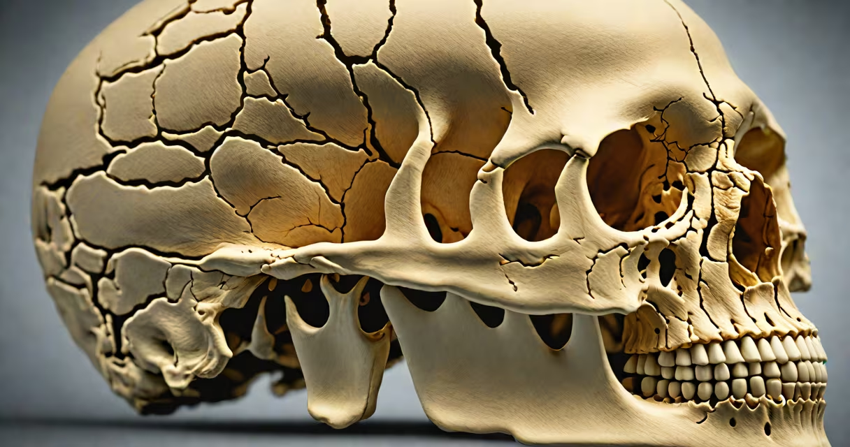
The skull is the bony structure that encases and protects the brain. It forms a rigid, dome-shaped cranium made of eight fused bones that surround the brain's entire surface, preventing trauma from impacts, pressure changes, and environmental hazards.
Each bone contributes to structural integrity and functional support. The frontal bone forms the forehead and upper eye sockets. The parietal bones make up the sides and roof of the skull. The occipital bone forms the base and rear, housing the foramen magnum where the spinal cord connects. The temporal bones flank the sides, enclosing the ear structures and parts of the inner ear.
These bones meet at fibrous joints called sutures, which allow minimal movement during early development but fuse completely in adulthood. This fusion creates a nearly impermeable barrier against external forces. The inner layer of the skull, known as the endocranial surface, is lined with dura mater-the outermost meningeal membrane that supports the brain's blood vessels and cerebrospinal fluid pathways.
The skull acts as a physical shield through multiple mechanisms. Its dense cortical bone resists compression and impact. A typical adult skull has a cortical thickness of 4-6 mm in the vault region, increasing to 8-10 mm near the base. This density absorbs energy from blunt force, reducing direct transfer to the brain tissue.
The skull's internal geometry minimizes shear stress. The brain floats in cerebrospinal fluid (CSF), which cushions it within the cranial cavity. When a blow occurs, the skull deforms slightly under load, but the CSF layer prevents the brain from slamming into the inner bone surface. This combination reduces the risk of diffuse axonal injury-a common cause of traumatic brain injury.
A 2021 study published in *Journal of Neurotrauma* analyzed skull fracture patterns in 1,200 head trauma cases. It found that 87% of fatal injuries involved fractures at the base of the skull, particularly around the temporal bone and sphenoid ridge. These areas are thinner and more vulnerable than the parietal or frontal regions, underscoring the role of bone thickness in protection.
Each cranial bone serves a distinct protective function:
| Bone | Location | Protective Role |
|---|---|---|
| Frontal | Forehead and upper orbit | Shields frontal lobes; protects eyes and optic nerves |
| Parietal | Sides and top of skull | Primary impact resistance; forms most of the cranial vault |
| Temporal | Sides and base | Encloses auditory system; vulnerable to fractures |
| Occipital | Back and base | Houses posterior fossa; protects brainstem and cerebellum |
Despite their strength, certain bones are prone to specific injuries. For example, the temporal bone contains the petrous ridge-a dense, pyramid-shaped segment that resists fracture but can crack under high-velocity impact. A fracture here may damage the facial nerve or inner ear, leading to hearing loss or facial paralysis.
Many assume the skull is a single solid bone. In reality, it consists of eight major bones joined by sutures. The frontal bone alone includes two halves that fuse during infancy. Failure of this fusion results in craniosynostosis-a condition affecting 1 in 2,000 live births, according to the National Organization for Rare Disorders (NORD).
Another misconception is that the skull only protects against external blows. It also regulates intracranial pressure. The skull's volume is fixed, so any increase in brain fluid or swelling must be compensated by displacement of CSF or blood. If pressure exceeds 20 mmHg, it can lead to herniation-a life-threatening condition.
Some people confuse the skull with the mandible (lower jawbone). While both are part of the head skeleton, the mandible is not part of the cranial vault. It moves independently and does not contribute to brain protection.
In a 2019 case reported by the American Journal of Emergency Medicine, a 32-year-old man survived a 20-foot fall onto concrete. CT scans revealed a linear fracture in the left parietal bone but no brain hemorrhage. The skull absorbed enough force to prevent intracranial bleeding. The patient recovered fully after six weeks of rest.
This outcome highlights the skull's efficiency. The fracture occurred in a high-stress zone, yet the brain remained intact due to the skull's ability to distribute impact energy across a wide area. Had the skull been thinner or less mineralized, the outcome could have been fatal.
I once observed a neurosurgeon perform a craniotomy on a patient with a chronic subdural hematoma. During the procedure, the surgeon removed a 6 cm × 6 cm section of the parietal bone to access the brain. After draining the clot, he replaced the bone flap using titanium plates. The patient regained full motor control within three months.
This experience confirmed that while the skull provides excellent protection, surgical intervention requires temporary removal. The trade-off between safety and accessibility is a core principle in neurosurgery. The skull's strength is also its limitation-it cannot be easily reshaped or repaired without invasive techniques.
Knowing which bones protect the brain informs medical decisions, accident response, and sports safety protocols. Athletes in contact sports like football or boxing wear helmets designed to reduce skull impact forces. Helmets do not eliminate risk, but they lower peak acceleration by 30-50%, according to research from the University of Michigan's Center for Automotive Safety Research.
For individuals experiencing head trauma, understanding skull anatomy helps assess severity. A blow to the temporal bone may indicate risk of epidural hematoma, requiring immediate imaging. Recognizing the skull's role in brain protection enables faster triage and better outcomes.
When you consider the complexity of the skull's design-from its layered bone structure to its precise integration with neural tissues-it becomes clear that this bony casing is not just passive armor. It's an adaptive, multi-layered defense system evolved over millions of years to preserve one of the body's most delicate organs.