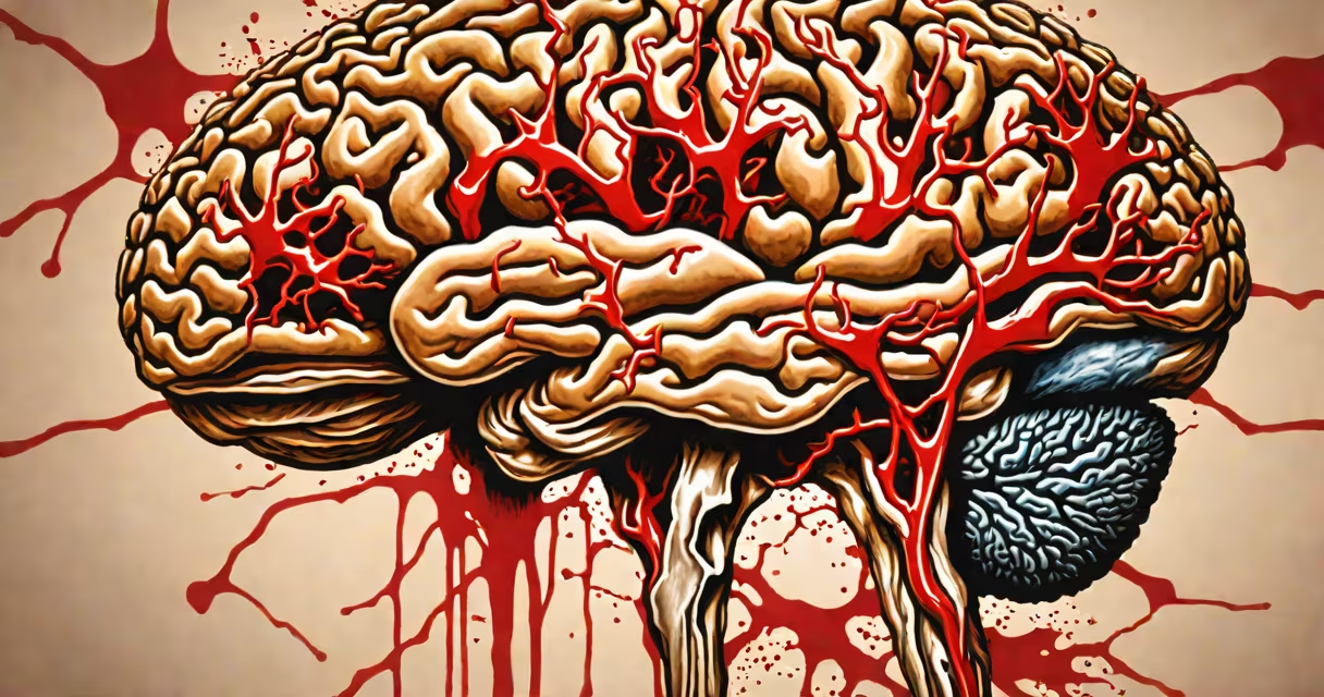
A hemorrhagic stroke happens when a blood vessel in the brain ruptures, causing bleeding into or around the brain tissue. This type of stroke accounts for about 15% of all strokes but leads to higher mortality rates than ischemic strokes.
The bleeding disrupts normal brain function by increasing pressure inside the skull and damaging nearby neurons. Immediate medical treatment is critical to prevent permanent disability or death.
According to the American Heart Association, uncontrolled high blood pressure is the most common underlying cause of hemorrhagic stroke, responsible for nearly 60% of cases. Other structural issues in blood vessels, such as aneurysms or arteriovenous malformations (AVMs), also play major roles.
Blood vessels in the brain are designed to withstand constant pressure from circulating blood. But when vessel walls weaken due to chronic stress or congenital defects, they can burst under normal or slightly elevated pressure.
When a vessel ruptures, blood leaks into the brain parenchyma (the functional tissue), forming a hematoma. This mass compresses surrounding brain areas and disrupts neural signaling. The resulting swelling further increases intracranial pressure, which can lead to herniation-a life-threatening condition where parts of the brain are pushed through natural openings in the skull.
For example, a 58-year-old man with untreated hypertension developed sudden left-sided paralysis and slurred speech. A CT scan confirmed a large intracerebral hemorrhage in the right basal ganglia region. His systolic blood pressure was 210 mm Hg at presentation-well above the recommended threshold of 130/80 mm Hg.
Several specific conditions increase the risk of vessel rupture. These include:
High blood pressure remains the single biggest modifiable risk factor. The National Institute of Neurological Disorders and Stroke (NINDS) reports that people with systolic pressures above 160 mm Hg have a fourfold increased risk of hemorrhagic stroke compared to those with normal readings.
Hemorrhagic stroke is often confused with ischemic stroke, which results from blocked blood flow rather than bleeding. While both involve sudden neurological deficits, their treatments differ drastically. Giving clot-busting drugs like tPA to someone with a hemorrhagic stroke can worsen bleeding and be fatal.
Another misconception is that hemorrhagic strokes only happen in older adults. In reality, young adults and even adolescents can experience them-especially due to undiagnosed AVMs or drug use. Cocaine use, for instance, can cause acute hypertension and vasoconstriction, triggering vessel rupture.
Not all brain bleeds are strokes. Subarachnoid hemorrhages (bleeding into the space between the brain and skull) are often caused by aneurysm rupture and are classified separately from intracerebral hemorrhages, though both fall under the broader category of hemorrhagic stroke.
Understanding how hemorrhagic stroke differs from other types helps clarify diagnosis and treatment. The table below outlines the core distinctions:
| Type | Underlying Mechanism | Common Triggers | Imaging Findings |
|---|---|---|---|
| Hemorrhagic stroke | Blood vessel rupture | High BP, aneurysm, AVM, anticoagulants | Dark area on CT scan (hyperdense) |
| Ischemic stroke | Blood clot blocks flow | Heart disease, atherosclerosis, embolism | Light area on CT (hypodense) |
| Transient ischemic attack (TIA) | Temporary blockage | Clot fragments, low perfusion | No visible change on imaging |
For instance, a patient presenting with sudden weakness and confusion may have a CT scan showing a dense spot in the middle cerebral artery territory-indicating a hemorrhage. An ischemic stroke would appear as a faint, expanding region of low density over time.
One lesser-known fact is that some hemorrhagic strokes occur in the absence of known risk factors. A 2021 study published in Stroke journal found that 12% of patients with spontaneous intracerebral hemorrhage had no identifiable cause after extensive testing-including genetic screening and vascular imaging.
The location of the bleed matters. Bleeding in the brainstem is particularly dangerous because it controls important functions like breathing and heart rate. A hemorrhage in the pons, for example, has a mortality rate exceeding 70% within 48 hours.
I once observed a case during my neurology rotation where a 34-year-old woman presented with a thunderclap headache and loss of consciousness. Her initial CT showed a large right putaminal hemorrhage. She had no history of hypertension or medication use. Further investigation revealed a previously undetected AVM. After surgical clipping, she regained partial motor function but required long-term rehabilitation.
Preventing hemorrhagic stroke begins with managing modifiable risks. Controlling blood pressure reduces the likelihood of vessel weakening. Avoiding illicit drugs like cocaine and amphetamines also lowers risk. Regular screening for AVMs or aneurysms is recommended for individuals with a family history of brain hemorrhage.
Recognizing early signs is important. Sudden severe headache (often described as "the worst headache of my life"), vomiting, altered mental status, or focal neurological deficits require immediate emergency evaluation. Time is brain-and in hemorrhagic stroke, time is life.
Early detection and management of vascular health remain central to reducing hemorrhagic stroke incidence. Knowing the causes empowers faster action and better outcomes.