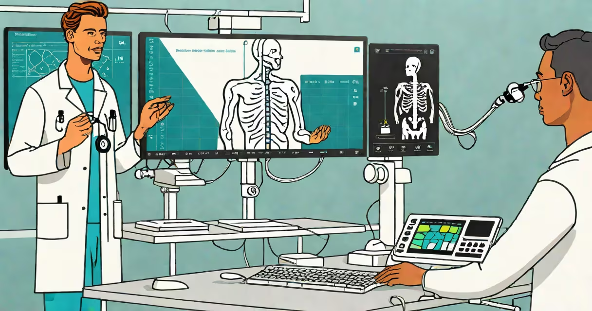
To become a registered diagnostic medical sonographer, you must complete a formal education program that includes specific science, anatomy, and clinical practice courses. These programs typically lead to a certificate, associate's degree, or bachelor's degree in sonography.
Ultrasound tech programs blend classroom instruction with hands-on clinical rotations. Students learn how to operate imaging equipment, interpret scans, and communicate findings to physicians. The curriculum is designed around national standards set by the American Registry of Diagnostic Medical Sonographers (ARDMS).
Coursework covers human anatomy, physiology, and pathology, along with physics principles governing sound wave behavior in tissue. Students also study patient safety, ethics, and medical terminology. Programs require completion of at least 1,000 hours of supervised clinical experience before certification eligibility.
Most accredited ultrasound tech programs require foundational science courses. These include biology, chemistry, and physics. A solid grasp of cellular structure, organ systems, and fluid dynamics is essential for understanding how ultrasound waves interact with tissues.
For example, a typical program includes:
These courses build the scientific foundation needed to interpret scan results. Without understanding how blood flows through vessels or how tumors appear on echocardiograms, sonographers cannot detect abnormalities accurately.
The Association of Medical Imaging Management (AMIM) reports that 92% of accredited programs include a dedicated course in ultrasound physics and instrumentation. This class teaches how transducers generate and receive sound waves, how frequency affects resolution, and how artifacts arise from improper technique.
Beyond general science, students take advanced courses tailored to their chosen specialty. Common specializations include abdominal, obstetric/gynecologic, vascular, and cardiac sonography.
Each track includes targeted coursework. For instance, abdominal sonography requires detailed study of the liver, kidneys, pancreas, and spleen. Students learn to identify conditions like fatty liver disease, renal cysts, and gallstones using grayscale and Doppler imaging.
| Specialty | Key Course Topics | Typical Credit Hours |
|---|---|---|
| Abdominal | Liver function, portal vein flow, kidney anatomy | 6 |
| OB/GYN | Fetal development stages, placental positioning, amniotic fluid volume | 5 |
| Vascular | Arterial stenosis, venous insufficiency, Doppler waveform analysis | 7 |
| Cardiac | Left ventricular ejection fraction, valvular regurgitation, pericardial effusion | 8 |
Students often complete a capstone project involving real patient cases under faculty supervision. One graduate from a program in Texas documented a rare case of a splenic artery aneurysm during her clinical rotation, which was later published in a regional radiology journal.
Every accredited program mandates clinical experience. Students rotate through hospitals, outpatient clinics, and specialty centers to perform scans under direct supervision. These rotations ensure competence in patient handling, equipment operation, and emergency response.
Rotations typically span 12 to 18 weeks and cover multiple specialties. A student may spend four weeks in obstetrics, four in abdominal imaging, and four in vascular labs. Each site uses different equipment models, exposing learners to varied software interfaces and machine settings.
During my time in a cardiac sonography rotation at a VA hospital, I performed over 150 echocardiograms. I learned to adjust gain settings in real time to improve endocardial borders and recognize subtle signs of left ventricular hypertrophy. The feedback from supervising sonographers helped me improve image quality and reduce repeat scans.
Many assume that ultrasound tech training is purely technical-just learning to press buttons. That's incorrect. The role demands clinical judgment, emotional intelligence, and critical thinking.
For example, a patient with suspected deep vein thrombosis may have swelling and pain. The sonographer doesn't just scan the leg veins-they assess the patient's history, review risk factors, and decide whether to perform a compression study or refer for further testing. This decision-making process is taught in clinical reasoning courses.
Another misconception is that all ultrasound techs work in hospitals. In reality, many work in private practices, mobile imaging services, or research institutions. The educational path remains similar, but elective courses may differ based on setting. A sonographer working in a sports medicine clinic might take additional courses in musculoskeletal imaging.
After completing an accredited program, graduates are eligible to sit for the ARDMS certification exam. Passing this exam allows entry into the workforce as a Registered Diagnostic Medical Sonographer (RDMS).
According to the U.S. Bureau of Labor Statistics, employment of diagnostic medical sonographers is projected to grow 12% from 2022 to 2032-faster than average. Demand is driven by aging populations and increased use of non-invasive imaging in early diagnosis.
Programs vary in duration. Certificate programs take 12 to 18 months. Associate's degrees take two years. Bachelor's programs take four years and include general education requirements. Students should verify accreditation status through the Commission on Accreditation of Allied Health Education Programs (CAAHEP).