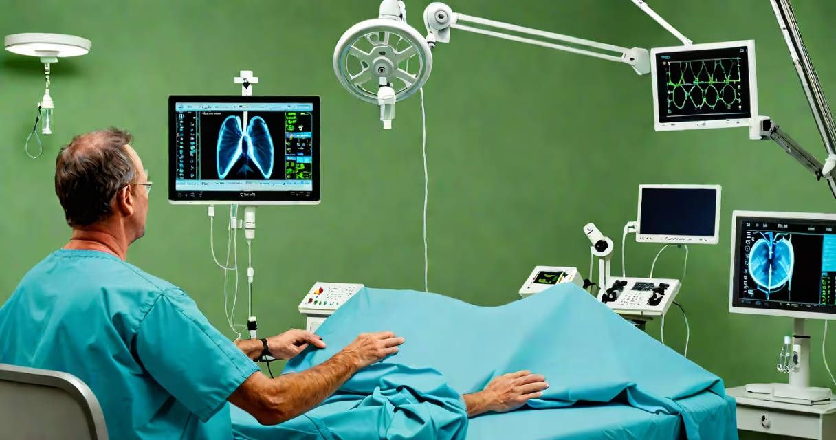
Cardiac catheterization is a minimally invasive medical procedure that involves inserting a thin, flexible tube (catheter) into a blood vessel, typically in the groin or wrist, and guiding it to the heart to assess heart function, measure pressures, and visualize coronary arteries using contrast dye and X-ray imaging.
This procedure is primarily used to diagnose coronary artery disease, evaluate heart valve function, and assess the severity of blockages in the coronary arteries. It is performed under local anesthesia and takes approximately 30 to 60 minutes, though recovery time varies based on the patient's condition and access site.
According to the American Heart Association, cardiac catheterization is one of the most common diagnostic procedures for patients with suspected heart disease, with over 1 million procedures performed annually in the United States. The technique allows physicians to make informed decisions about treatment, including whether to proceed with angioplasty or bypass surgery.
The procedure begins with a small incision at the femoral artery in the upper thigh or the radial artery in the wrist. A guidewire is inserted through a sheath, followed by the catheter, which is advanced under fluoroscopic guidance toward the heart. Once positioned, contrast dye is injected to show blood flow through the coronary arteries.
Real-time X-ray images (angiograms) capture the movement of the dye, revealing areas of narrowing or blockage. Pressure measurements within the heart chambers can also be taken to assess blood flow dynamics and determine if surgical intervention is needed.
A 2021 study published in the Journal of the American College of Cardiology found that cardiac catheterization accurately identifies significant coronary artery stenosis (≥70% narrowing) in 94% of cases, making it a gold standard for pre-intervention assessment.
There are several types of cardiac catheterization, each tailored to specific clinical goals:
| Type | Purpose | Common Use Cases |
|---|---|---|
| Diagnostic Coronary Angiography | Visualize coronary arteries | Assessing chest pain, evaluating heart attack risk |
| Left Heart Catheterization | Measure pressures in left heart chambers | Evaluating heart failure, mitral valve function |
| Right Heart Catheterization | Measure pulmonary artery pressure | Detecting pulmonary hypertension, assessing right-sided heart strain |
| Interventional Catheterization | Treat blockages during the procedure | Performing balloon angioplasty, placing stents |
While diagnostic catheterization focuses solely on evaluation, interventional catheterization includes therapeutic steps such as stent placement. The latter is often referred to as percutaneous coronary intervention (PCI).
One non-obvious distinction is that radial access (wrist entry) is now preferred in many centers due to lower bleeding risk and faster recovery. A 2020 meta-analysis in *The Lancet* showed radial access reduced major bleeding events by 34% compared to femoral access, especially in high-risk patients.
Cardiac catheterization is conducted in specialized cardiac catheterization labs within hospitals or outpatient cardiac centers. These rooms are equipped with real-time imaging systems, hemodynamic monitoring, and emergency resuscitation tools.
It is recommended for patients with stable angina, acute chest pain, abnormal stress test results, or those being evaluated before heart surgery. It is also used in patients who have had a heart attack to determine if immediate revascularization is necessary.
For example, a 62-year-old man with persistent chest pain despite medication underwent cardiac catheterization via radial access. The procedure revealed a 90% blockage in the left anterior descending artery. He was treated with a drug-eluting stent during the same session, restoring blood flow and relieving symptoms.
Many people confuse cardiac catheterization with open-heart surgery. Unlike surgery, it does not require a sternotomy or general anesthesia. The procedure is done under local anesthetic, and most patients remain awake but sedated.
Another misconception is that it is inherently dangerous. While complications occur in less than 1% of cases, risks include bleeding at the access site, allergic reaction to contrast dye, kidney injury, and rare arterial damage. However, the procedure is considered safe when performed by experienced interventional cardiologists.
Contrary to popular belief, cardiac catheterization is not always followed by stenting. In some cases, the findings may support medical management instead. A 2022 report from the National Cardiovascular Data Registry indicated that nearly 40% of diagnostic catheterizations led to no further intervention, highlighting the role of the procedure in avoiding unnecessary procedures.
I once observed a catheterization in progress where a patient developed mild hypotension during dye injection. The team quickly stopped the infusion, administered fluids, and stabilized the patient within five minutes. This reinforced how critical real-time monitoring is during the procedure.
After the procedure, patients are monitored for 1 to 2 hours. They must lie flat to prevent bleeding at the access site. Most patients can go home the same day, especially if radial access was used.
Patients should avoid heavy lifting and strenuous activity for 24 hours. Follow-up care includes reviewing results with a cardiologist and discussing treatment options. If stents were placed, dual antiplatelet therapy (e.g., aspirin and clopidogrel) is prescribed for at least one year to prevent clotting.
Cardiac catheterization remains a key part of modern cardiology. Its ability to provide precise anatomical and functional data makes it indispensable in managing heart disease.