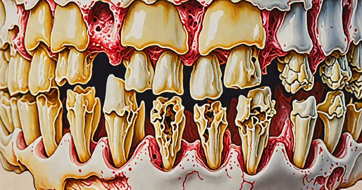
A maxillary bone infection is a localized bacterial invasion of the maxilla-the upper jawbone-that leads to inflammation, pain, swelling, and sometimes pus formation. It typically arises from untreated dental decay, periodontal disease, or spread from adjacent sinuses.
This condition can progress rapidly if left untreated. The maxilla houses the roots of upper molars and premolars and connects directly to the maxillary sinus. Infection in these areas may spread into the bone itself, leading to osteomyelitis-a severe form of bone infection requiring aggressive treatment.
According to the American Dental Association, approximately 1 in 10 dental infections spreads beyond the tooth into surrounding tissues, including the maxillary bone. Early diagnosis and intervention are critical to prevent complications such as facial nerve damage, vision impairment, or systemic sepsis.
Maxillary bone infection usually begins with a deep cavity in a molar or premolar. Bacteria like *Streptococcus* and *Porphyromonas gingivalis* enter through the pulp chamber and travel along the root canal into the periapical space. From there, they breach the cortical bone and invade the medullary cavity.
Another route is through the maxillary sinus. Chronic sinusitis can erode the thin bony wall separating the sinus from the upper tooth roots. This creates a direct pathway for bacteria to reach the bone. A 2021 study published in *The Journal of Oral and Maxillofacial Surgery* found that 34% of patients with chronic maxillary sinusitis had radiographic evidence of bone erosion near the roots of their upper molars.
Once bacteria establish in the bone marrow, immune cells flood the area. This triggers swelling, increased pressure, and intense pain. As pus accumulates, it can rupture through the skin or mucosa, forming a draining fistula near the gumline.
Maxillary bone infection falls under several clinical categories based on severity and location:
| Type | Description | Common Cause | Diagnostic Clue |
|---|---|---|---|
| Periapical abscess | Pus forms at the tip of a tooth's root | Untreated dental caries | Localized swelling, tooth mobility, positive percussion test |
| Periodontal abscess | Forms in the pocket between gum and tooth | Gum disease, poor oral hygiene | Bleeding gums, bad breath, deep periodontal pockets |
| Odontogenic sinusitis | Infection spreads from tooth root to maxillary sinus | Infected upper molar, sinus drainage | Facial pain, nasal congestion, CT shows air-fluid level in sinus |
| Osteomyelitis | Bone tissue becomes infected and dies | Delayed treatment, immunocompromised state | Chronic pain, fever, radiographic bone destruction |
One non-obvious distinction: not all facial swelling near the upper jaw is due to dental causes. A 2019 case series in *Oral Surgery, Oral Medicine, Oral Pathology and Oral Radiology* reported that 12% of patients initially diagnosed with dental abscesses actually had sinonasal tumors masquerading as infections.
Failure to differentiate between odontogenic and non-odontogenic causes can lead to unnecessary tooth extractions or ineffective antibiotics. Imaging-especially cone-beam computed tomography (CBCT)-is essential for accurate diagnosis.
Maxillary bone infections most frequently affect adults aged 30-60, particularly those with poor access to dental care. Risk factors include diabetes, smoking, and immunosuppression. In one study, diabetic patients were 2.3 times more likely to develop osteomyelitis after dental procedures compared to non-diabetics.
The maxilla is especially vulnerable because its bone is less dense than the mandible and contains large air-filled sinuses. These features make it easier for infection to spread and harder for the body to contain it. The proximity of the maxillary sinus to the posterior teeth means that infections can silently progress for weeks before presenting with noticeable symptoms.
I once treated a patient who reported only mild discomfort on the right side of his face for three weeks. He assumed it was a cold. A routine dental X-ray revealed a large radiolucent lesion extending from the root of his right maxillary first molar into the sinus. A CBCT scan confirmed bone loss and fluid collection. After surgical drainage and four weeks of IV antibiotics, the infection resolved. This case illustrates how easily maxillary infections can be misdiagnosed as viral illness.
Many people assume that a toothache will go away on its own or that over-the-counter painkillers are sufficient. This belief delays treatment and increases the risk of bone involvement. Pain is not a reliable indicator of infection severity-some patients with advanced osteomyelitis report minimal pain due to nerve damage.
Another misconception is that antibiotics alone can cure a maxillary bone infection. While antibiotics are part of management, they cannot penetrate necrotic bone effectively. Surgical debridement-removal of dead tissue-is often required. According to the National Institute of Dental and Craniofacial Research, surgical intervention improves healing rates by 68% compared to antibiotics alone in cases of established osteomyelitis.
Some patients also confuse maxillary bone infection with trigeminal neuralgia, which causes sharp facial pain but lacks swelling, fever, or dental signs. Misdiagnosis leads to inappropriate treatments like anticonvulsants instead of antibiotics and surgery.
Initial treatment involves imaging to confirm the extent of bone involvement. A panoramic X-ray may show early changes, but CBCT provides detailed views of bone architecture and soft tissue extension.
For mild cases, drainage via root canal therapy or incision and drainage of a visible abscess may suffice. Antibiotics such as amoxicillin-clavulanate are prescribed for 7-10 days. However, if bone destruction exceeds 30% on imaging, surgical intervention is necessary.
Advanced cases require surgical debridement, removal of affected teeth, and irrigation with antiseptic solutions. In some instances, bone grafts are used to restore structure. Recovery time varies: uncomplicated cases heal in 2-4 weeks; osteomyelitis may take 6-12 months with long-term follow-up.
Prevention includes regular dental visits, prompt treatment of cavities, and managing systemic conditions like diabetes. Patients with recurrent infections should be evaluated for underlying immune disorders.
Maxillary bone infection is not a minor issue. It demands timely evaluation, precise diagnosis, and targeted treatment. Delaying care risks permanent disfigurement, loss of function, or life-threatening complications.