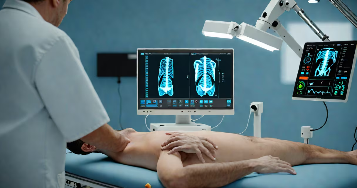
Ultrasound technicians, also known as diagnostic medical sonographers, use high-frequency sound waves to produce real-time images of internal body structures. They operate specialized equipment to visualize organs, blood flow, and fetal development without radiation exposure. Their work supports diagnosis, monitors treatment progress, and guides minimally invasive procedures.
These professionals work under physician supervision but independently perform most scan operations. They position patients, apply gel to improve signal transmission, adjust machine settings for optimal clarity, and identify anatomical features during live imaging. After capturing images, they review them for quality and report findings to doctors.
According to the U.S. Bureau of Labor Statistics, ultrasound techs held about 96,000 jobs in 2022, with demand projected to grow 17% through 2032-faster than average. This growth stems from aging populations needing more diagnostic imaging and increasing use of ultrasound in prenatal care and cardiology.
Ultrasound machines emit pulses of sound waves above human hearing range (typically 2-18 MHz). These waves travel into the body and bounce off tissues at different rates based on density. The returning echoes are captured by the transducer, converted into electrical signals, and processed into visual images on a screen.
Technicians manipulate the probe manually to change angles and focus points. For example, in abdominal scans, they may tilt the probe to view the liver from multiple planes. In obstetrics, they track fetal movement and measure limb lengths to estimate gestational age. Each scan requires precise positioning and timing to avoid artifacts like shadowing or reverberation.
A single scan session can last 15 to 45 minutes depending on complexity. A cardiac ultrasound (echocardiogram) may require recording heart motion across several heartbeats to assess valve function. Technicians must maintain steady hand control and monitor image quality throughout.
Most ultrasound techs specialize in one area. The most common fields include:
| Specialty | Primary Use Cases | Typical Patient Population |
|---|---|---|
| Abdominal | Liver disease, gallstones, kidney cysts | Adults with abdominal pain or elevated liver enzymes |
| Obstetric/Gynecologic | Prenatal screening, fetal anomalies, ovarian cysts | Pregnant women, adolescents with pelvic pain |
| Cardiac (Echocardiography) | Heart valve function, ejection fraction, congenital defects | Patients with chest pain, heart failure, or suspected cardiomyopathy |
| Vascular | Deep vein thrombosis, carotid stenosis, aneurysms | Stroke risk assessment, post-surgical monitoring |
Each specialty demands distinct skills. Obstetric sonographers must recognize fetal anatomy and detect developmental issues early. Vascular techs analyze blood flow patterns using Doppler ultrasound to identify blockages. Cardiac sonographers interpret complex heart motions and calculate parameters like stroke volume.
For example, during a routine 20-week prenatal ultrasound, a technician identifies a ventricular septal defect-a hole in the baby's heart wall-using 2D and color Doppler imaging. The finding is documented and reported to the referring obstetrician, who then arranges follow-up testing.
Many assume ultrasound techs simply press a button and read results. In reality, they make real-time decisions based on anatomy, patient history, and image quality. A technician may alter frequency or gain settings mid-scan to clarify a suspicious mass in the pancreas.
Another misconception is that ultrasound replaces MRI or CT scans. While ultrasound excels in soft tissue visualization and is safe for pregnant women, it cannot penetrate bone or air-filled spaces well. It's often used alongside other modalities rather than as a standalone substitute.
Ultrasound techs do not diagnose diseases. They provide images and measurements that physicians interpret. A 2021 study published in *Radiology* found that sonographers correctly identified 94% of clinically significant abnormalities during routine screening, but final diagnoses were made by radiologists or specialists.
Becoming an ultrasound tech typically requires completing an accredited program lasting 12 to 24 months. Programs offer associate degrees or certificates. Coursework includes anatomy, physiology, physics of sound waves, patient communication, and clinical rotations.
After graduation, technologists must pass a certification exam administered by the American Registry for Diagnostic Medical Sonography (ARDMS). Certification is not mandatory in all states but is widely expected by employers. As of 2023, over 85% of employed sonographers held ARDMS credentials.
I once observed a student struggle to identify the portal vein in a liver scan due to poor angle placement. After adjusting the probe and repositioning the patient, the structure became visible. That moment highlighted how technical skill and hands-on practice matter more than theoretical knowledge alone.
For those interested in entry-level roles, what classes do you need for ultrasound tech outlines foundational subjects like biology, math, and medical terminology. Advanced training may include echocardiography or vascular certification.
Ultrasound techs integrate into larger healthcare teams. They communicate findings verbally and in written reports. A typical workflow begins with a physician's referral, followed by scheduling, patient intake, scan execution, and result delivery.
They also manage patient concerns. A woman undergoing a first-trimester scan may worry about fetal viability. The technician explains the process, shows the heartbeat on screen, and reassures her while maintaining professionalism.
Some facilities use AI-assisted tools to flag potential abnormalities. However, these systems still require human validation. A 2022 pilot at Johns Hopkins Hospital showed that combining AI detection with sonographer review reduced missed findings by 30% compared to manual review alone.
When a patient arrives with acute abdominal pain, the ultrasound tech performs a focused assessment with sonography in trauma (FAST) exam. They quickly scan for free fluid around the liver and spleen-indicating internal bleeding. Results are relayed within minutes to emergency staff.