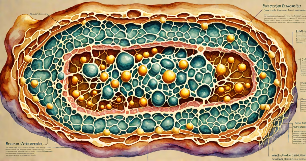
The endoplasmic reticulum (ER) is a continuous membrane-bound organelle in eukaryotic cells that forms a network of flattened sacs and tubules. It functions in protein folding, lipid synthesis, and calcium storage, with two main structural forms: rough ER and smooth ER.
The endoplasmic reticulum originates from the outer membrane of the nuclear envelope and extends throughout the cytoplasm. Its structure consists of interconnected cisternae (flattened sacs) and anastomosing tubules. These membranes are composed of phospholipid bilayers with embedded proteins. The lumen-the space inside the ER-is continuous with the perinuclear space surrounding the nucleus.
Electron microscopy reveals that the rough ER has ribosomes attached to its cytosolic surface, giving it a granular appearance. In contrast, the smooth ER lacks ribosomes and appears smoother under the microscope. This structural difference directly determines function: rough ER synthesizes transmembrane and secretory proteins, while smooth ER handles lipid metabolism and detoxification.
The rough ER's ribosome-studded surface enables co-translational protein insertion. As a polypeptide chain emerges from a ribosome, a signal recognition particle binds to a signal sequence on the nascent protein. This complex docks onto a receptor in the ER membrane, allowing the growing chain to be threaded into the lumen. Once inside, chaperone proteins like BiP assist in proper folding. Misfolded proteins are retrotranslocated to the cytosol for degradation via the ubiquitin-proteasome system.
The smooth ER specializes in lipid synthesis and ion regulation. In liver cells, smooth ER contains enzymes such as cytochrome P450, which metabolize drugs and toxins. In steroid-producing cells like those in the adrenal cortex, smooth ER synthesizes cholesterol-derived hormones. The tubular geometry increases surface area for enzyme activity and allows efficient diffusion of lipids and ions.
A single hepatocyte contains approximately 20,000 to 30,000 smooth ER tubules. These structures form a changing network that remodels rapidly in response to metabolic demands. For example, alcohol consumption induces smooth ER proliferation in liver cells-a compensatory mechanism to improve detoxification capacity.
There are two primary structural subtypes of the endoplasmic reticulum, each with specialized morphology and biochemical composition:
| Type | Structure | Key Components | Primary Functions |
|---|---|---|---|
| Rough ER | Flattened cisternae with ribosomes bound to the cytosolic side | Signal recognition particle (SRP), SRP receptor, Sec61 translocon | Protein synthesis, folding, glycosylation, quality control |
| Smooth ER | Tubular network; no ribosomes | Cytochrome P450, glucose-6-phosphatase, Ca²⁺-ATPase | Lipid synthesis, drug detoxification, calcium storage |
Both types are continuous with each other and with the nuclear envelope. The transition zone between rough and smooth regions contains specific proteins like reticulons and DP1, which stabilize membrane curvature and regulate domain identity.
Recent cryo-electron tomography studies show that the smooth ER forms highly curved tubules with diameters averaging 50-80 nm. These tubules are stabilized by BAR-domain proteins that sense and induce membrane bending. This structural adaptation maximizes surface-to-volume ratio for enzymatic reactions.
Many assume the endoplasmic reticulum is a static structure. In reality, it is highly changing. Membrane domains constantly exchange components through vesicular transport and membrane fusion events. The ER network can reorganize within minutes in response to stress signals, such as unfolded protein accumulation.
Another misconception is that all protein synthesis occurs on the rough ER. While most secretory and membrane proteins are synthesized there, some cytosolic proteins are translated on free ribosomes in the cytoplasm. The presence of a signal peptide determines whether a protein enters the ER.
Some confuse the ER with the Golgi apparatus. Both are involved in protein modification, but they differ structurally and functionally. The ER synthesizes and folds proteins; the Golgi modifies them further and sorts them for delivery. The ER is a single continuous compartment; the Golgi consists of stacked cisternae.
Consider cystic fibrosis, caused by mutations in the CFTR gene. The most common mutation, ΔF508, results in a misfolded CFTR protein. This protein fails to pass quality control in the rough ER and is degraded before reaching the plasma membrane. As a result, chloride transport is impaired, leading to thick mucus in lungs and pancreas.
Studies using live-cell imaging show that mutant CFTR accumulates in the ER lumen and triggers the unfolded protein response (UPR). This activates transcription factors like ATF6 and IRE1α, which upregulate chaperones and ER-associated degradation (ERAD) machinery. Without proper ER structure and function, cellular homeostasis collapses.
I observed this process firsthand during a lab experiment using HeLa cells expressing fluorescently tagged CFTR. After inducing stress with tunicamycin (an N-glycosylation inhibitor), we tracked ER morphology changes via confocal microscopy. The rough ER expanded visibly, and misfolded proteins aggregated in clusters-visual evidence of structural strain.
Disruptions in ER structure correlate with several diseases. Alzheimer's disease involves abnormal accumulation of amyloid precursor protein (APP) fragments in the ER lumen. Parkinson's disease features α-synuclein aggregates that impair ER-Golgi trafficking. Cancer cells often exhibit enlarged, fragmented ER networks to support high rates of protein synthesis.
Research from the National Institutes of Health confirms that mutations in genes encoding ER-shaping proteins (e.g., Reticulon 1, REEP1) cause hereditary spastic paraplegia. These conditions show how structural integrity of the endoplasmic reticulum is essential for neuronal health.
Understanding the structure of endoplasmic reticulum explains how cells maintain proteostasis. Its dual architecture-rough and smooth-enables specialization. Without this organization, protein folding, lipid production, and calcium signaling would fail, disrupting cellular life.