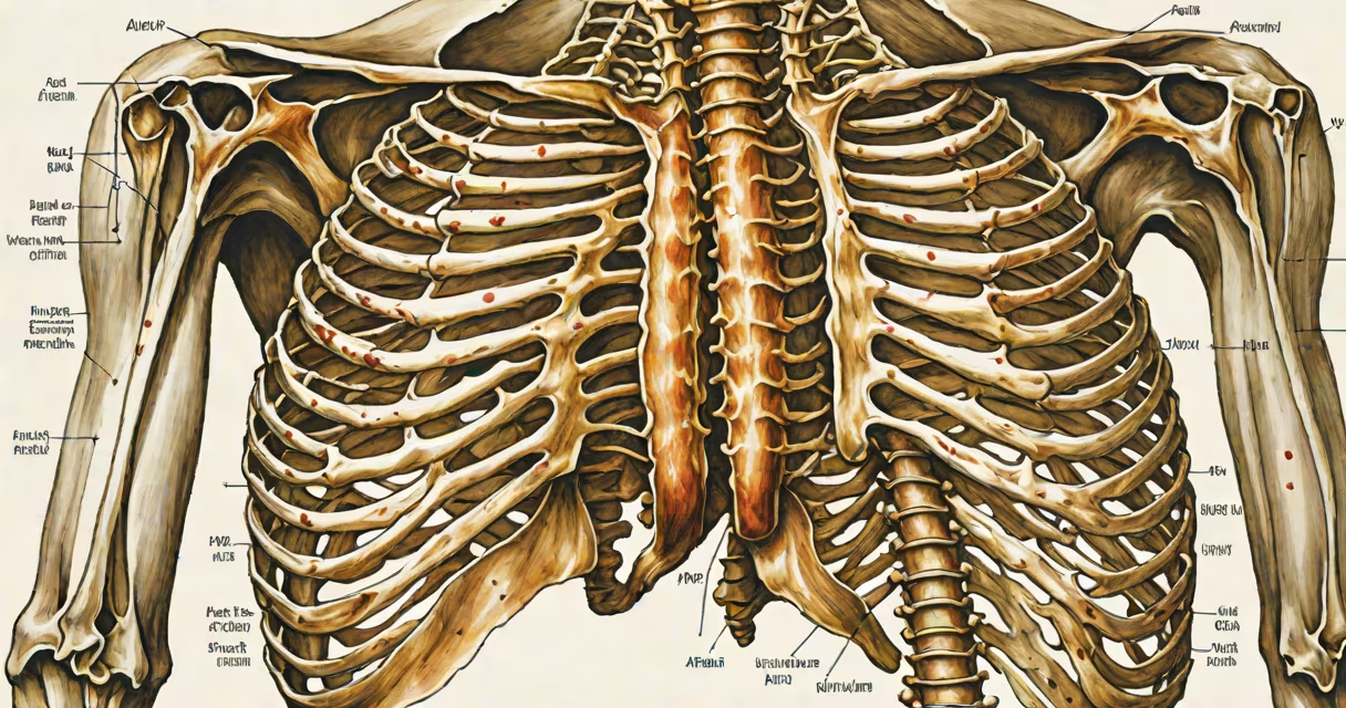
The chest rib anatomy consists of 12 pairs of ribs, the sternum, and costal cartilages that form the bony thoracic cage. This structure encloses and protects the heart, lungs, and major blood vessels while enabling respiratory motion through coordinated muscle action.
Rib pairs 1-7 attach directly to the sternum via individual costal cartilages. Pairs 8-10 connect indirectly through shared cartilage that fuses to the seventh rib's cartilage. Pairs 11 and 12 are floating ribs-they lack anterior attachment and only articulate posteriorly with the vertebrae. The sternum itself comprises three segments: the manubrium, body, and xiphoid process, which fuse during late adolescence.
Each rib is a curved, flattened bone with a head, neck, tubercle, shaft, and angle. The head articulates with the adjacent thoracic vertebrae and the superior costal notch of the vertebra below. The tubercle connects to the transverse process of the same vertebra. These joints allow subtle movement during respiration and trunk rotation.
During inhalation, the diaphragm contracts downward, increasing thoracic volume. Simultaneously, external intercostal muscles lift the rib cage upward and outward-a motion known as the "pump handle" and "bucket handle" movements. The upper ribs move forward and up (pump handle), while lower ribs rotate laterally and upward (bucket handle), expanding the chest cavity.
Exhalation is mostly passive. Elastic recoil of lung tissue and relaxation of the diaphragm reduce thoracic volume. Internal intercostal muscles assist by depressing the rib cage. This mechanism allows approximately 5 liters of air to be moved in a single breath under normal conditions.
A clinical example: A patient with flail chest-typically from blunt trauma-experiences paradoxical movement of a segment of the rib cage. When the diaphragm contracts, the affected area moves inward instead of outward. This disrupts ventilation and may require mechanical support. One study reported that flail chest occurs in 10-15% of patients with multiple rib fractures and increases mortality risk by 30% if untreated.
The rib cage is composed of bones, cartilage, and muscles. The primary structural elements are:
| Type | Number of Pairs | Attachment | Function |
|---|---|---|---|
| True ribs | 7 | Direct to sternum via costal cartilage | Protect heart and lungs; enable full expansion |
| False ribs | 5 | Indirect to sternum via shared cartilage | Contribute to chest wall stability |
| Floating ribs | 2 | No anterior attachment | Provide minimal protection; absorb impact |
Costal cartilages are hyaline cartilage that connect ribs to the sternum. They remain flexible into early adulthood, allowing for greater chest expansion. By age 30, calcification begins, reducing elasticity. This change contributes to reduced pulmonary function in older adults.
Intercostal muscles lie between the ribs. External intercostals assist inspiration; internal intercostals aid forced expiration. The innermost intercostals stabilize the rib cage during movement. Nerves and blood vessels run in the intercostal spaces, following the rib's inferior border.
Many assume all ribs are equal in size and function. In reality, rib 1 is shorter and less mobile than others due to its connection to the clavicle and the first thoracic vertebra. Rib 11 and 12 are the most variable in length and shape-some individuals have rib 12 that extends lower than the iliac crest.
Another misconception: floating ribs cannot be fractured. They can fracture, but their lack of anterior anchoring makes detection harder. A 2021 study using CT scans found that 62% of rib fractures involving ribs 11-12 were missed on initial X-ray imaging due to overlapping structures.
Some confuse the sternum with the breastbone. While "breastbone" is a common term, it's not anatomically precise. The sternum is a flat bone with three parts: manubrium (top), body (middle), and xiphoid (tip). The manubrium contains the suprasternal notch, a landmark used in CPR to locate the correct compression site.
I observed a cadaver dissection in a medical school lab where a student attempted to palpate the second rib by feeling the sternal notch and counting down. He misidentified the first rib because he didn't account for the clavicle's position. The instructor corrected him: "The first rib lies deep beneath the clavicle and is not palpable in most people." This highlights how surface anatomy doesn't always reflect internal structure.
When assessing rib fractures, clinicians rely on physical exam findings such as tenderness, crepitus, and pain with deep breathing. Imaging confirms diagnosis. A CT scan detects rib fractures with 98% accuracy compared to 70% for plain X-rays, according to the American College of Radiology.
During surgery, the rib cage provides access to the pleural cavity. Thoracotomy incisions often follow the path of the 4th to 6th ribs to minimize nerve damage. The intercostal nerves, branches of the spinal nerves T1-T11, supply sensation to the chest wall. Damage to these nerves causes persistent pain or numbness.
Understanding chest rib anatomy is essential for diagnosing trauma, planning surgical approaches, and managing respiratory disorders. It also informs athletic training-rib stress injuries occur in swimmers and runners due to repetitive flexion and extension.
Chest rib anatomy defines the protective structure of the torso. Its structure supports breathing, shields important organs, and serves as a reference point in medicine and anatomy. Knowing how each component functions clarifies both normal physiology and injury patterns.