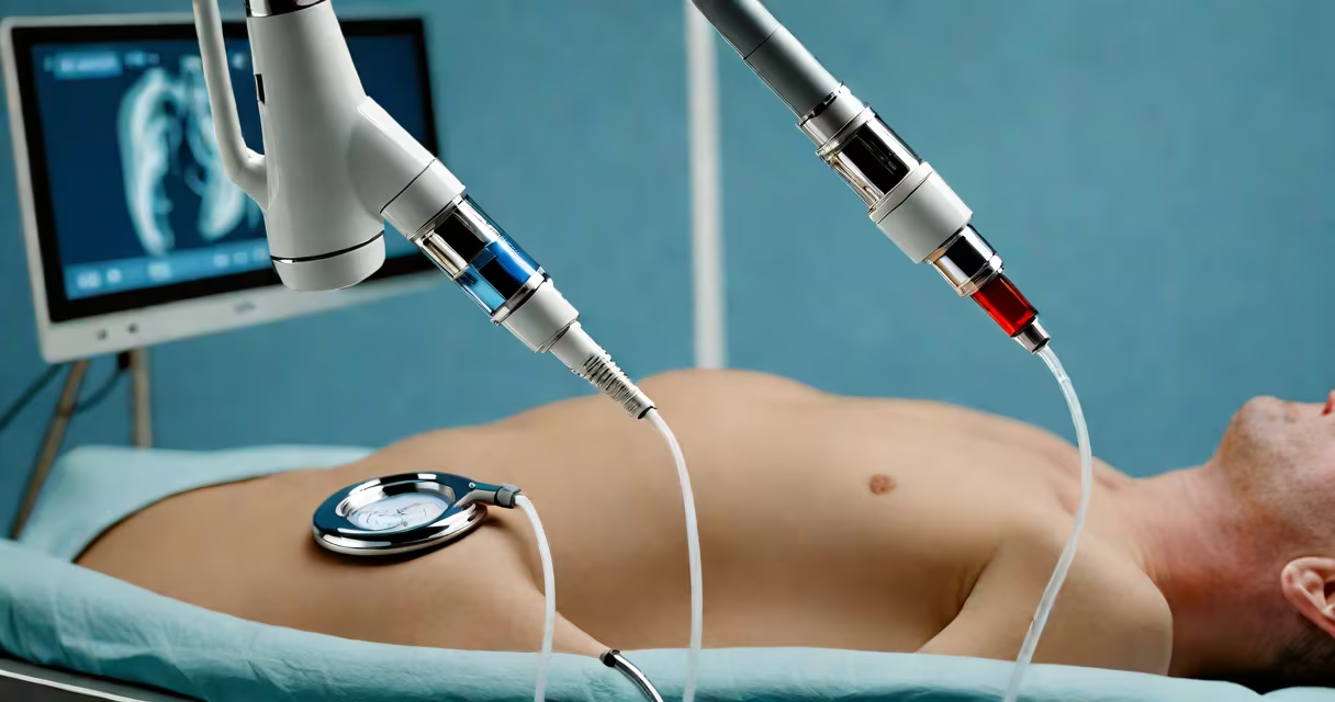
The tee vs tte procedure refers to two distinct cardiac imaging techniques: transesophageal echocardiography (TEE) and transthoracic echocardiography (TTE). TTE uses a handheld probe on the chest wall to capture heart images, while TEE involves inserting a probe into the esophagus behind the heart for higher-resolution views. Both assess heart structure and function, but TEE offers clearer detail of posterior structures like the left atrial appendage and aortic root.
TTE is performed with the patient lying on their back or left side. A gel is applied to the chest, and a technician moves the probe across several chest positions-apical, parasternal, subcostal-to obtain multiple views. The entire process takes 20 to 30 minutes and requires no sedation. TTE is widely available and used for initial evaluation of heart failure, valve disease, and congenital defects.
TEE requires moderate sedation and is done in a cardiac cath lab or dedicated echo set. The probe is inserted through the mouth, guided down the esophagus, and positioned near the heart. Because the esophagus lies directly behind the heart, TEE provides superior resolution of the posterior chambers, mitral valve, and interatrial septum. It is often used when TTE images are suboptimal due to obesity, lung disease, or poor acoustic windows.
TTE relies on ultrasound waves reflected off moving heart tissues. The probe emits sound waves at frequencies between 2 and 5 megahertz. These waves bounce off heart walls, valves, and blood flow, returning to the probe as echoes. A computer translates the time and intensity of each echo into a real-time image of heart motion and chamber size. Doppler mode measures blood velocity across valves to detect regurgitation or stenosis.
TEE uses a similar principle but with a smaller, rotating transducer mounted on a flexible tube. The probe is advanced until it reaches the lower esophagus, approximately 20 cm from the mouth. Once in place, the transducer rotates 360 degrees, capturing cross-sectional images of the heart from multiple angles. This allows visualization of complex anatomy such as patent foramen ovale (PFO), thrombi in the left atrial appendage, and aortic dissection.
A 2021 study published in *JACC: Cardiovascular Imaging* found that TEE detected PFO in 87% of patients with cryptogenic stroke, compared to 53% with TTE. The improved sensitivity stems from reduced tissue interference and proximity to the heart. TEE also reduces false-negative rates in evaluating prosthetic valve function.
Both procedures serve different purposes based on clinical context. TTE is the first-line test for suspected heart failure, valvular disease, or acute chest pain. It is non-invasive, low-cost, and widely accessible. TTE is used in emergency departments to screen for pulmonary embolism using the McConnell sign-a pattern of right ventricular dysfunction.
TEE is reserved for specific scenarios where TTE fails to provide adequate images or when high diagnostic certainty is needed. Common indications include:
Table: Key Differences Between TTE and TEE
| Feature | Transthoracic (TTE) | Transesophageal (TEE) |
|---|---|---|
| Probe Location | Chest wall | Esophagus |
| Sedation Required | No | Yes (moderate) |
| Image Quality (Posterior Structures) | Low to moderate | High |
| Procedure Time | 20-30 min | 30-45 min |
| Complication Risk | Very low | Minor (e.g., sore throat, rare esophageal injury) |
One non-obvious distinction: TTE cannot reliably visualize the left atrial appendage unless the patient is in a specific position. TEE, however, captures a clear view of the appendage in 98% of cases, making it essential for assessing stroke risk in atrial fibrillation patients.
Many assume TEE is simply a "better" version of TTE. That's incorrect. TTE remains the standard screening tool because it's faster, safer, and sufficient for most conditions. TEE is not interchangeable-it's complementary. Using TEE routinely increases cost and exposure without improving outcomes in low-risk populations.
Another misconception is that TEE is painful. While insertion causes temporary discomfort, patients receive conscious sedation. A 2020 review in *Circulation: Cardiovascular Imaging* reported that 94% of patients rated TEE comfort as "acceptable" or better, with only 2% reporting significant distress.
I once observed a patient with a history of rheumatic fever who had a normal TTE but was later diagnosed with a mobile thrombus in the left atrium via TEE. The TTE missed it due to poor window caused by chronic lung disease. This case underscored that TEE isn't optional in certain high-risk groups-it's necessary.
Consider a 68-year-old man admitted with sudden-onset left-sided weakness. CT scan shows no hemorrhage. TTE reveals normal ejection fraction but a small, mobile mass in the left atrium. The TTE image is blurry due to emphysema. A TEE confirms a 1.2 cm thrombus in the left atrial appendage. The diagnosis changes management: anticoagulation is initiated, and a neurologist is consulted for stroke prevention.
Another example: a 45-year-old woman with recurrent migraines and a family history of stroke undergoes TTE, which shows no abnormalities. Her doctor orders a TEE to rule out PFO. The TEE identifies a patent foramen ovale with right-to-left shunt. She undergoes device closure and experiences fewer migraines post-procedure.
These cases show that TTE is effective for broad screening, but TEE delivers precision where it matters most. The American Society of Echocardiography recommends TEE when TTE results are inconclusive or when high-risk conditions are suspected.