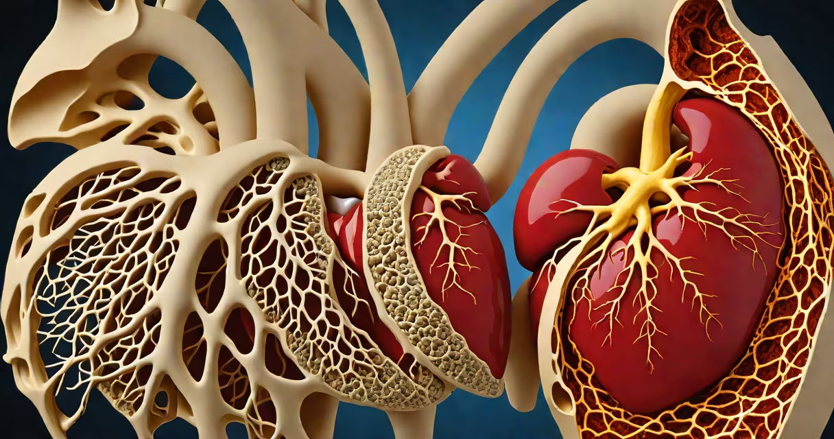
Fluid build up around the heart, medically termed pericardial effusion, occurs when excess fluid accumulates in the pericardial space-the area between the heart and its protective sac. This condition may develop slowly or rapidly and can impair heart function if pressure increases.
The pericardium is a double-layered sac that surrounds the heart. Normally, a small amount of fluid lubricates the space between the layers. When this balance is disrupted, fluid can accumulate due to injury, inflammation, or disease. The most common causes include infections, autoimmune disorders, trauma, and underlying heart conditions.
According to the American Heart Association, pericardial effusion affects approximately 1 in 1,000 hospital admissions and is often linked to systemic illnesses. The Mayo Clinic notes that in up to 30% of cases, no clear cause is identified-termed "idiopathic" effusions-but they still require monitoring.
Fluid enters the pericardial space through increased permeability of blood vessels or leakage from damaged tissues. The process begins when the pericardium becomes inflamed (pericarditis), causing fluid to seep into the space. Alternatively, fluid may leak from blood vessels due to high pressure, such as in heart failure or after surgery.
When fluid builds up quickly, it compresses the heart-a condition called cardiac tamponade. This reduces the heart's ability to fill with blood, leading to low blood pressure, rapid breathing, and fainting. The condition requires emergency treatment. A 2021 study in *Circulation* found that tamponade develops in about 15% of large effusions, emphasizing the need for early detection.
Chronic effusions grow slowly over weeks or months. These often result from long-term conditions like kidney failure or cancer. The body may adapt to the extra volume, delaying symptoms until significant pressure builds.
Pericardial effusion arises from several distinct pathways. The most frequent causes include:
A 2020 review in *The Lancet* highlighted that tuberculosis remains a leading infectious cause in developing regions, accounting for nearly 40% of cases in sub-Saharan Africa. In contrast, viral pericarditis dominates in North America and Western Europe, with enteroviruses responsible for over half of diagnosed cases.
Effusions are categorized by size and speed of development. Small effusions may cause no symptoms and resolve on their own. Large or rapidly accumulating effusions pose immediate risks.
| Type | Size | Onset | Common Causes | Risk Level |
|---|---|---|---|---|
| Small | <10 mm | Slow | Idiopathic, mild infection | Low |
| Moderate | 10-20 mm | Gradual | Heart failure, hypothyroidism | Moderate |
| Large | >20 mm | Acute | Trauma, post-op, cancer | High |
Not all large effusions are dangerous. Some patients tolerate large volumes without hemodynamic compromise. However, sudden onset-even with moderate fluid-can signal tamponade. The presence of pulsus paradoxus (a drop in blood pressure during inspiration) is a clinical red flag.
Pericardial effusion is frequently mistaken for other cardiac issues. It shares symptoms with heart failure, pulmonary embolism, and pleural effusion. Distinguishing features include the location of fluid and associated signs.
For example, pleural effusion involves fluid around the lungs, not the heart. Patients with pleural effusion typically have shortness of breath and decreased breath sounds on one side. Pericardial effusion may show muffled heart sounds and distant heart tones on exam.
A key diagnostic clue is echocardiography. A standard transthoracic echo can detect fluid behind the heart and assess for tamponade. A 2019 study in *JAMA Cardiology* found that echo sensitivity for detecting effusion exceeds 95%, making it the gold standard.
A 62-year-old man presented with fatigue and swelling in his legs. He had a history of stage III lung cancer. An echocardiogram revealed a 25-mm effusion with signs of tamponade: elevated jugular venous pressure, narrow pulse pressure, and pulsus paradoxus. The fluid was drained via pericardiocentesis, and analysis confirmed malignant cells.
This case illustrates that cancer-related effusions can be massive and life-threatening. The patient required ongoing oncology care alongside cardiology management. Without drainage, he would have progressed to cardiogenic shock.
Fluid build up around the heart is not always due to serious illness. Some people develop small effusions after minor chest trauma or viral illness. But any new onset of chest pain, shortness of breath, or unexplained fatigue warrants evaluation.
Seek immediate medical attention if you experience chest tightness, dizziness, or fainting. These may indicate tamponade. Even subtle symptoms like persistent fatigue or swollen ankles should prompt a visit to a doctor.
One non-obvious fact: effusions can occur in healthy individuals during intense physical activity. A 2022 study in *European Heart Journal* documented transient pericardial fluid in 12% of endurance athletes post-marathon. The fluid resolved within 48 hours without intervention-proof that temporary fluid isn't always pathological.
Fluid build up around the heart is a sign, not a diagnosis. Identifying the root cause determines treatment. Early imaging and clinical assessment prevent complications. If you suspect this condition, consult a cardiologist promptly.