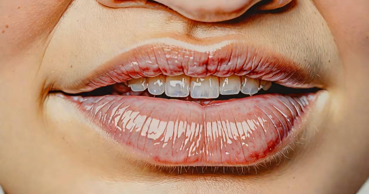
A mucocele is a small, fluid-filled cyst that forms in the mouth, typically appearing as a soft, painless, translucent or bluish swelling. It develops when a minor injury damages a salivary gland duct, causing saliva to leak into surrounding tissue. Most commonly found on the inner surface of the lower lip, mucoceles are usually less than 1 cm in diameter and may fluctuate in size.
They resemble a tiny bubble or blister under the skin, often with a smooth surface and a waxy or gelatinous texture. The color ranges from clear to pale pink or deep blue, depending on the amount of trapped fluid and blood. Unlike a cold sore, which forms a crusty lesion near the lips, a mucocele sits flat against the mucosa and doesn't ulcerate.
According to the American Academy of Oral and Maxillofacial Pathology, mucoceles account for nearly 45% of all minor salivary gland lesions in the oral cavity. They are most common in children and young adults, likely due to increased lip biting or chewing habits during development.
Mucoceles arise when a salivary gland duct becomes blocked or ruptures. Common triggers include accidental biting, tongue thrusting, or dental appliance irritation. Once saliva leaks into the connective tissue beneath the mucous membrane, the body responds by forming a cystic sac to contain the fluid.
The most frequent site is the lower lip, especially the vermilion border-the area between the red lip and the skin. Other locations include the floor of the mouth (known as a ranula), the inner cheeks, and the tongue's underside. A ranula is larger and deeper, sometimes extending into the neck region, and may require surgical drainage.
When examining a mucocele, it feels soft and compressible. Pressing gently may cause it to shrink temporarily as fluid shifts, then refill. This characteristic distinguishes it from solid growths like fibromas or tumors.
While all mucoceles share core features, their appearance varies based on location and duration:
| Type | Location | Appearance | Duration |
|---|---|---|---|
| Simple mucocele | Lower lip, inner cheek | Clear or bluish, 2-6 mm, soft | Days to weeks; may resolve spontaneously |
| Ranula | Floor of mouth | Translucent, dome-shaped, up to 2 cm | Persistent; may recur if untreated |
| Deep mucocele | Submucosal tissue | Less visible, firm, may mimic tumor | Longer duration; harder to diagnose clinically |
People often mistake mucoceles for cold sores, aphthous ulcers, or even early signs of oral cancer. Cold sores appear as grouped vesicles that crust over and heal within 7-10 days. Aphthous ulcers are painful, shallow, round sores with a yellow center and red border. Oral cancer lesions are typically persistent, irregular, and may bleed easily.
A key difference: mucoceles are not painful unless irritated. They don't develop a central ulceration. If a bump lasts longer than two weeks, changes color, bleeds, or grows rapidly, it should be evaluated by a dentist or oral surgeon.
In a 2020 study published in the Journal of Oral Pathology & Medicine, researchers analyzed 127 cases of mucoceles in patients aged 5-35. Of those, 89% were located on the lower lip, and 72% resolved within six weeks without treatment. However, 18% recurred after initial rupture, suggesting that trauma prevention is key.
I once observed a 10-year-old patient who had a 5-mm bluish bump on the inner lip that had been present for three weeks. The child reported no pain and no history of trauma. On examination, the lesion was compressible and translucent. After confirming it was a mucocele via clinical diagnosis, we advised monitoring. Two weeks later, the child returned with the lesion gone-likely due to spontaneous rupture and reabsorption of fluid.
This case highlights a lesser-known fact: mucoceles can disappear on their own after bursting. The fluid drains, the tissue heals, and the cyst collapses. But recurrence is common, especially in individuals prone to lip biting.
Most mucoceles are harmless and resolve independently. However, consult a healthcare provider if the bump:
If removal is needed, a minor surgical procedure called marsupialization or excision under local anesthesia is effective. These procedures have high success rates and low complication rates, according to the American Dental Association.
Other conditions that mimic mucoceles include epidermoid cysts, hemangiomas, and salivary gland tumors. A biopsy may be required to rule out malignancy in rare cases, particularly in adults over 40.
Understanding what a mucocele looks like helps avoid unnecessary anxiety. It's not a sign of serious illness. But knowing when to act ensures timely care and prevents complications.