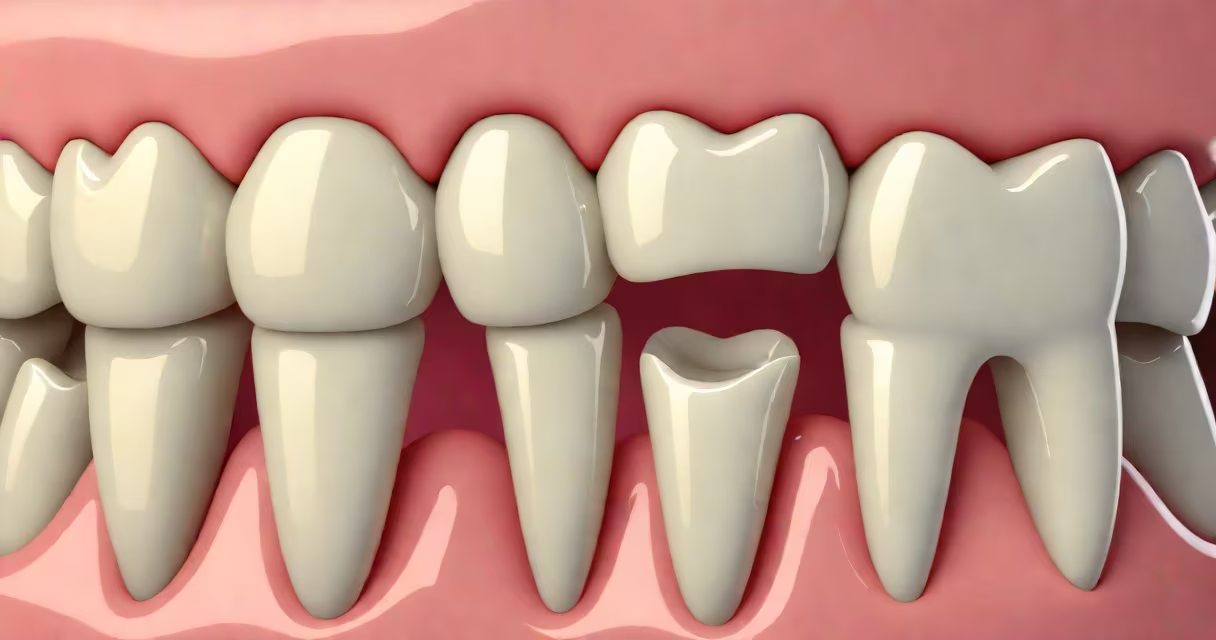
White bumps on gum line are small, raised areas of tissue that appear near the base of teeth, often indicating a localized issue such as a dental cyst, torus, or infection. They may be painless or tender, and their presence signals a need for clinical evaluation to rule out serious conditions.
These bumps form when fluid, cells, or debris accumulate beneath the gum surface. They typically appear as firm, whitish nodules that may grow slowly or remain stable in size. While some are harmless, others signal underlying pathology requiring treatment. Ignoring them risks progression to abscesses or bone loss.
According to the American Dental Association (ADA), persistent oral lesions-especially those lasting more than two weeks-should be evaluated by a dentist or oral surgeon. Early diagnosis prevents complications like tooth mobility or systemic infection.
White bumps on gum line arise from trapped material or abnormal tissue growth. The most common cause is a dentigerous or radicular cyst, which forms around a tooth's root tip due to inflammation from untreated decay or trauma. These cysts fill with fluid and expand slowly, pushing gum tissue upward.
Another mechanism involves hyperkeratosis-excessive buildup of keratin in the skin layer. This can result from chronic irritation, such as from ill-fitting dentures or aggressive brushing. The keratin hardens into a small, white nodule visible at the gum margin.
A third possibility is a torus, a bony growth that develops on the jawbone. Though usually asymptomatic, tori can project above the gum line and appear as white or pinkish lumps. They're benign and genetically influenced, often discovered during routine X-rays.
Differentiating between types of white bumps on gum line requires visual and clinical assessment. Below is a comparison of common origins:
| Type | Appearance | Texture | Common Location | Underlying Cause |
|---|---|---|---|---|
| Dental cyst | Smooth, dome-shaped, white or yellowish | Firm, sometimes fluctuant | At root tip, near molar or premolar | Chronic infection or dead nerve |
| Hyperkeratotic lesion | Flat or slightly raised, white or gray | Hard, rough to touch | Along gumline, especially lower front teeth | Repetitive friction or pressure |
| Torus mandibularis | Round or oval, white or pinkish | Very hard, bony | Lower jaw, near tongue side | Genetic bony overgrowth |
Each type responds differently to treatment. For example, a dental cyst may require surgical drainage or root canal therapy, while a hyperkeratotic bump may resolve with removal of the irritant.
Any white bump on gum line that persists beyond 14 days, increases in size, bleeds, or causes discomfort should be examined. Pain, swelling, or discharge suggest infection. A dentist may take a radiograph to assess whether the bump is connected to a tooth root or bone structure.
I once observed a patient with a 6mm white bump near a molar that had been present for six weeks. It was painless but slightly movable. An X-ray revealed a periapical cyst. After surgical removal and root canal treatment, the lesion resolved completely within eight weeks. The patient reported no recurrence after two years.
The Mayo Clinic notes that untreated dental cysts can erode surrounding bone and lead to tooth loss. Early intervention reduces risk and preserves function.
People often mistake white bumps on gum line for canker sores or cold sores. Canker sores are shallow ulcers inside the mouth, not raised. Cold sores occur on lips and are caused by herpes simplex virus. Neither appears on the gum line as a firm, white nodule.
Another confusion arises with leukoplakia-a precancerous condition marked by thickened white patches. Unlike white bumps on gum line, leukoplakia is flat, cannot be scraped off, and is linked to tobacco or alcohol use. It requires biopsy to rule out dysplasia.
White bumps on gum line are not the same as gingivitis. Gingivitis causes redness, swelling, and bleeding at the gumline-not white nodules. The absence of redness or swelling helps distinguish these conditions.
Diagnosis begins with a visual exam and palpation. If the bump is firm and immobile, imaging is essential. A cone beam CT scan or panoramic X-ray shows internal structure and relationship to adjacent teeth and bone.
Treatment depends on the cause. A dental cyst may be treated with a minor surgical procedure called enucleation, where the cyst lining is removed. Root canal therapy may be needed if the tooth's pulp is infected. Hyperkeratotic lesions often improve when the source of friction-like a sharp edge on a filling-is smoothed.
If the bump is a torus, no treatment is needed unless it interferes with denture fitting or causes irritation. In rare cases, surgical removal is performed for cosmetic reasons.
For persistent or uncertain cases, referral to an oral surgeon is standard. The American Academy of Oral and Maxillofacial Radiology recommends imaging for any intraoral lesion suspected of being cystic or neoplastic.
Maintaining good oral hygiene reduces the risk of infection-related bumps. Brush twice daily with a soft-bristled brush, floss daily, and avoid chewing on hard objects like ice or pens. Regular dental checkups catch early signs of decay or gum disease.
If you notice a white bump, do not attempt to squeeze or scrape it. This can introduce bacteria and worsen inflammation. Avoid spicy or acidic foods that may irritate the area. Use a gentle, fluoride mouthwash to reduce plaque buildup.
White bumps on gum line are not normal. Their presence indicates a biological response to stress, infection, or structural change. Identifying the cause through clinical evaluation ensures proper management and long-term oral health.