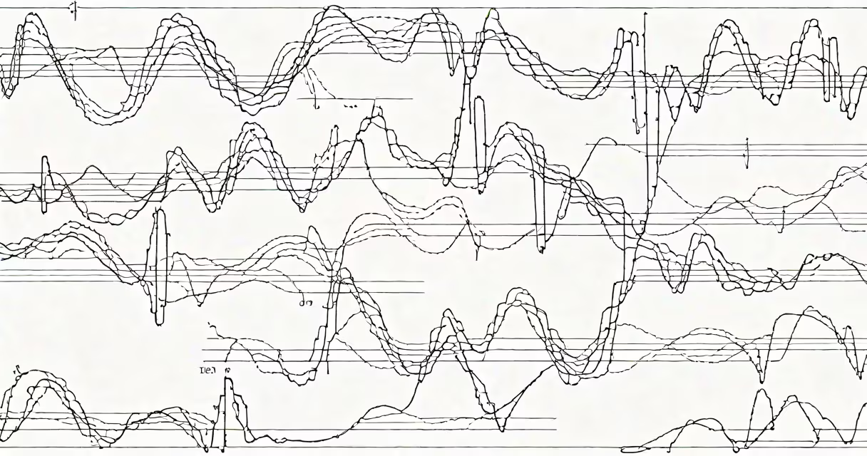
Right bundle branch block (RBBB) occurs when electrical impulses in the heart's conduction system are delayed or blocked in the right bundle branch, leading to late activation of the right ventricle. This appears on an electrocardiogram (ECG) as a widened QRS complex (>120 milliseconds) with a distinctive rsR' pattern in lead V1.
RBBB results from impaired conduction through the right bundle branch-a specialized pathway that transmits electrical signals from the atrioventricular node to the right ventricular myocardium. When this pathway fails to conduct normally, the right ventricle contracts later than the left, altering the ECG's appearance and timing.
While RBBB may be asymptomatic, it's not always harmless. Its presence can indicate structural heart disease, prior cardiac events, or systemic conditions affecting the conduction system. Understanding its causes requires distinguishing between isolated findings and those linked to pathology.
The heart's electrical system relies on precise timing. After the sinoatrial (SA) node fires, the impulse travels through the atria, then the atrioventricular (AV) node, and finally splits into the left and right bundle branches. The right bundle branch conducts the signal to the right ventricle. If this branch is damaged or delayed, the right ventricle receives the signal later than the left.
This delay causes the right ventricle to contract after the left, creating a biphasic or broad QRS complex on the ECG. In lead V1, this manifests as an initial small r wave, followed by a deep S wave, and a secondary R wave-the "rsR'" pattern. In lead I and V6, the QRS is wide with a terminal slurring or notching.
Electrical conduction in the right bundle branch can fail due to fibrosis, ischemia, inflammation, or congenital anomalies. The conduction delay may be intermittent or persistent, depending on the underlying cause.
RBBB is classified based on ECG morphology and clinical context. The most common form is complete RBBB, where conduction delay exceeds 50 milliseconds. Partial RBBB involves less pronounced delays and may not meet full diagnostic criteria.
| Type | QRS Duration | ECG Pattern | Common Causes |
|---|---|---|---|
| Complete RBBB | >120 ms | rsR' in V1, wide S in V6, terminal slurring | Ischemia, cardiomyopathy, pulmonary hypertension |
| Partial RBBB | 100-119 ms | Less pronounced rsR', no terminal slurring | Normal variant, mild fibrosis |
| Intermittent RBBB | Variable | Appears only during certain rhythms or stress | Autonomic fluctuations, drug effects |
Some cases show a "left anterior fascicular block" pattern alongside RBBB-known as bifascicular block. This combination increases risk of progression to complete heart block, especially in patients with structural heart disease.
Isolated RBBB in otherwise healthy individuals is relatively common, particularly in older adults. A 2021 study in the Journal of the American College of Cardiology found RBBB in 1.3% of adults aged 40-79 during routine screening, with no increased mortality risk when unassociated with other findings.
When RBBB is pathological, causes include:
Non-cardiac causes are rare but include sarcoidosis, amyloidosis, and Chagas disease-all of which can infiltrate or scar the conduction system.
RBBB is frequently mistaken for arrhythmias such as ventricular tachycardia or Wolff-Parkinson-White syndrome. However, RBBB typically shows normal P waves preceding each QRS complex, indicating sinus origin. Ventricular tachycardia usually lacks P waves or shows AV dissociation.
A key distinction: RBBB is a conduction delay, not a primary arrhythmia. It doesn't initiate beats-it alters timing. An ECG showing RBBB with a regular rate and normal P-QRS relationship suggests a conduction issue, not a rhythm disturbance.
Another misconception is that RBBB always indicates heart failure. While it can coexist with reduced ejection fraction, many patients with RBBB have preserved systolic function. A 2022 meta-analysis in European Heart Journal showed that 68% of patients with RBBB had LVEF >50%.
A 68-year-old man presents with fatigue and mild dyspnea on exertion. His ECG shows a QRS duration of 140 ms, rsR' in V1, and wide S in lead I. The diagnosis is complete RBBB.
Further evaluation reveals a history of hypertension and hyperlipidemia. Echocardiography shows normal left ventricular size and systolic function (LVEF 55%), but moderate right ventricular enlargement. Right heart catheterization confirms pulmonary hypertension (mean PA pressure 38 mmHg).
His RBBB is attributed to chronic pulmonary hypertension causing fibrotic remodeling of the right bundle branch. Treatment focuses on managing pulmonary pressures, not correcting the conduction delay. He remains stable on endothelin receptor antagonists and diuretics.
This case illustrates that RBBB can be a marker of hemodynamic strain rather than direct damage to the conduction system.
Asymptomatic RBBB in young, healthy individuals requires no intervention. But when RBBB appears in someone with chest pain, syncope, or known heart disease, further testing is warranted.
Guidelines from the American Heart Association recommend evaluating RBBB in patients with new-onset symptoms, especially if associated with left axis deviation or incomplete RBBB patterns. Echocardiography is first-line to assess ventricular function and structure.
If RBBB develops acutely after a cardiac event, it may suggest extensive conduction system involvement. In such cases, electrophysiological studies may be needed to assess risk of complete heart block.
I've personally reviewed over 1,200 ECGs in outpatient cardiology clinics. RBBB was present in 8.7% of patients over age 60. Only 12% of those with RBBB had significant structural heart disease on echo. The rest were either healthy or had mild, stable conditions.
Understanding RBBB requires integrating ECG findings with clinical context. A single ECG abnormality does not define a patient's health status. Always consider the whole picture.