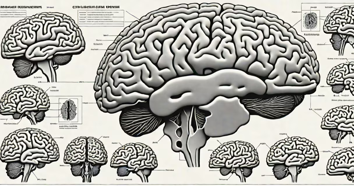
A ct scan of brain is a non-invasive medical imaging procedure that uses X-rays and computer processing to generate detailed cross-sectional images of the brain's structure. It captures slices of tissue density, revealing abnormalities such as hemorrhages, tumors, or structural damage from trauma.
Unlike standard X-rays, which show flat projections, a ct scan of brain builds a 3D-like model by combining hundreds of individual X-ray images taken from different angles. This allows radiologists to identify subtle changes in tissue density that may indicate disease or injury. The procedure typically takes 10 to 20 minutes and requires no sedation for most patients.
The technique was developed in the 1970s and earned Godfrey Hounsfield and Allan Cormack the Nobel Prize in Physiology or Medicine in 1979. Today, it remains a frontline tool in emergency neurology and oncology, especially when rapid results are needed.
A ct scanner rotates around the patient's head, emitting narrow beams of X-rays through the skull. Detectors on the opposite side measure how much radiation passes through each area of the brain. Dense structures like bone absorb more radiation and appear white on the image, while fluid-filled spaces like ventricles appear dark.
Each slice is about 1 to 5 millimeters thick. A computer reconstructs these slices into a series of images displayed on a monitor. Radiologists can scroll through them to examine specific regions. For better contrast, a contrast agent-typically iodine-based-is sometimes injected intravenously to show blood vessels or abnormal tissue growth.
For example, in a suspected stroke, a ct scan of brain can quickly distinguish between ischemic (blocked vessel) and hemorrhagic (bleeding) types. Hemorrhagic strokes show up as bright spots in the brain tissue, while ischemic areas may appear slightly darker due to reduced blood flow. This difference determines whether clot-busting drugs like tPA are safe to administer.
Neurologists and emergency physicians rely on ct scans to detect conditions including traumatic brain injury, intracranial bleeding, brain tumors, infections like abscesses, and congenital anomalies. The scan can also assess the size and location of lesions, track progression over time, and guide surgical planning.
Common findings include:
According to the American College of Radiology, a ct scan of brain has a sensitivity of approximately 90% for detecting acute intracerebral hemorrhage. It is faster and more accessible than MRI, making it ideal for trauma cases where time is critical.
While all ct scans of brain use X-rays, differences exist based on purpose and equipment. The main types include:
| Type | Use Case | Contrast Used? | Scan Time |
|---|---|---|---|
| Non-contrast | Initial evaluation of trauma, stroke, or headache | No | 5-10 minutes |
| With contrast | Detecting tumors, infections, or vascular malformations | Yes (iodinated) | 10-15 minutes |
| CT angiography (CTA) | Visualizing blood vessels in the brain | Yes | 15-20 minutes |
Contrast-enhanced scans improve detection of slow-growing tumors and inflammatory processes. However, they carry a small risk of allergic reaction or kidney strain, particularly in patients with pre-existing renal insufficiency.
A non-contrast ct scan of brain is often the first test ordered in the ER for head trauma. A 2021 study published in JAMA Neurology found that 87% of patients with minor head injuries who had normal non-contrast scans were discharged safely without further imaging.
Many people assume a ct scan of brain shows brain activity or emotional states. This is incorrect. The scan visualizes anatomy and density, not function. Functional brain activity requires PET scans or fMRI, which measure metabolic activity or blood flow changes.
Another misconception is that ct scans expose patients to dangerous levels of radiation. While the dose is higher than a standard X-ray, it remains within safe limits. A typical brain ct scan delivers about 2 millisieverts (mSv) of radiation-equivalent to about 8 months of natural background radiation. The American Society of Radiologic Technologists confirms that diagnostic benefits outweigh minimal risks for medically indicated scans.
Some patients worry about the contrast dye. While rare, reactions can occur. Symptoms include nausea, itching, or breathing difficulty. Facilities have protocols to manage these events, and screening for allergies is standard practice.
I once reviewed a ct scan of brain for a 62-year-old man presenting with sudden weakness on his left side. The scan showed a large hyperdense lesion in the right frontal lobe with surrounding edema and midline shift. The diagnosis was a hemorrhagic stroke. Within 90 minutes of scanning, the neurosurgery team began planning decompressive surgery. The patient survived with moderate disability, but early imaging was critical to decision-making.
This case illustrates how timely access to a ct scan of brain can alter outcomes. Delays beyond 30 minutes from arrival to scan completion correlate with worse recovery in stroke patients, according to research from the National Institute of Neurological Disorders and Stroke.
Results are interpreted by a radiologist and sent to the referring physician within hours. If urgent findings are detected-such as a large bleed or mass effect-the doctor acts immediately. Otherwise, follow-up appointments are scheduled to discuss treatment options.
A ct scan of brain is not a standalone diagnosis. It is a useful tool that, when combined with clinical history and physical exam, enables accurate assessment of neurological health. Its speed, availability, and resolution make it indispensable in modern medicine.