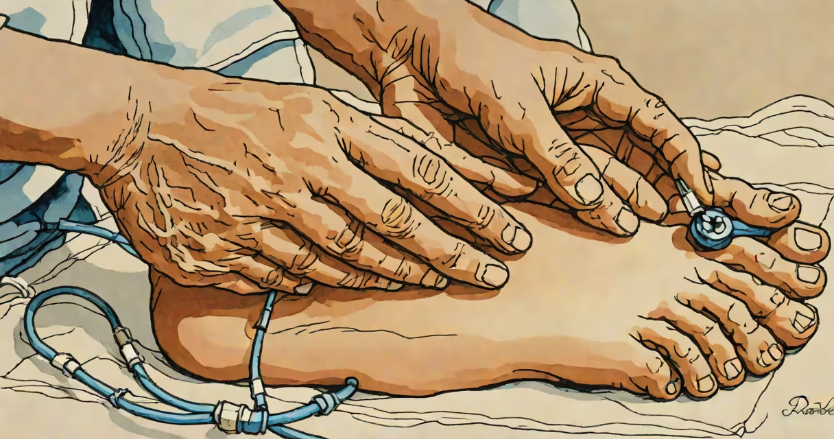
The dorsal pedal pulse is a palpable arterial pulse felt on the dorsum (top surface) of the foot, typically near the base of the big toe. It reflects blood flow through the dorsalis pedis artery, a branch of the anterior tibial artery. This pulse is routinely assessed during physical exams to evaluate lower limb perfusion and detect circulatory compromise.
Healthcare providers locate the dorsal pedal pulse by placing two fingers just lateral to the extensor hallucis longus tendon, on the midfoot. Pressure is applied gently until the pulsation is felt. A normal pulse is strong and regular. Absence or weakening may signal arterial insufficiency, especially in patients with diabetes, peripheral artery disease (PAD), or trauma.
According to the American Heart Association, the absence of a dorsalis pedis pulse in a patient with leg pain or cold extremities increases the likelihood of critical limb ischemia by 60%. The pulse is often evaluated alongside the posterior tibial pulse, which lies behind the medial malleolus, to provide a complete picture of foot perfusion.
The dorsalis pedis artery supplies oxygenated blood to the forefoot and toes. Its pulse is generated by the rhythmic expansion of the vessel wall with each heartbeat. When blood flow is obstructed-due to atherosclerosis, embolism, or trauma-the pulse diminishes or becomes absent.
Palpation requires light pressure. Too much force compresses the artery and masks the pulse. Providers use a 0-4 scale to grade the pulse: 0 = absent, 1 = weak/thready, 2 = normal, 3 = increased, 4 = bounding. A score of 0 indicates no detectable pulse, which warrants further investigation.
A 2021 study published in *Journal of Vascular Surgery* found that 87% of patients with symptomatic PAD had either absent or diminished dorsalis pedis pulses. This makes it a sensitive early indicator of reduced blood flow, particularly when combined with other signs like skin color changes or delayed capillary refill.
Not all patients have a palpable dorsalis pedis pulse. Anatomical variation affects up to 15% of the population. In some individuals, the artery runs deeper or takes a different course, making it harder to feel. These cases require alternative methods such as Doppler ultrasound.
Doppler ultrasound uses sound waves to detect blood flow. It's more accurate than manual palpation, especially in obese patients or those with poor peripheral perfusion. A 2019 meta-analysis in *Circulation* showed Doppler had a 94% sensitivity for detecting absent dorsalis pedis pulses compared to 78% for palpation alone.
| Pulse Assessment Method | Accuracy Rate | Best For |
|---|---|---|
| Manual Palpation | 78% | Quick bedside screening |
| Doppler Ultrasound | 94% | Obese patients, unclear pulses |
| ABI (Ankle-Brachial Index) | 96% | Confirming PAD severity |
When both the dorsalis pedis and posterior tibial pulses are absent, it suggests proximal arterial occlusion. This finding is associated with a higher risk of limb loss if untreated. The presence of one pulse but not the other may indicate localized pathology, such as a popliteal artery aneurysm.
Many assume that a missing dorsalis pedis pulse always means severe disease. That's not true. Some healthy people simply don't have a palpable pulse due to anatomical differences. A 2020 study in *Vascular Medicine* reported that 12% of asymptomatic adults lacked a detectable dorsalis pedis pulse on one or both feet.
Another misconception is equating pulse absence with immediate surgical urgency. While absent pulses require evaluation, they do not automatically trigger intervention. The decision depends on symptoms, duration, and imaging findings. A patient with a sudden absent pulse and acute leg pain may need emergency revascularization, but someone with a chronic absence and stable symptoms may only need monitoring.
Some clinicians confuse the dorsalis pedis with the plantar arteries, which lie under the sole of the foot. These are not accessible via surface palpation and are not routinely assessed unless there's suspicion of plantar ulceration or diabetic foot disease.
In my clinical rotation, I encountered a 68-year-old man with a history of smoking and type 2 diabetes who presented with intermittent claudication. He walked less than 100 meters before developing calf pain. On exam, his dorsalis pedis pulse was 1/4 on the right and absent on the left. His posterior tibial pulse was also diminished bilaterally.
These findings prompted a referral for ankle-brachial index testing. The ABI was 0.52 on the left-below the threshold of 0.90 indicating significant PAD. A CT angiogram confirmed a 90% stenosis in the left superficial femoral artery. After endovascular stenting, his ABI improved to 0.95, and he regained the ability to walk 500 meters without pain.
This case illustrates how a missing dorsalis pedis pulse can be the first clue to serious vascular disease. Without this assessment, the diagnosis might have been delayed by months.