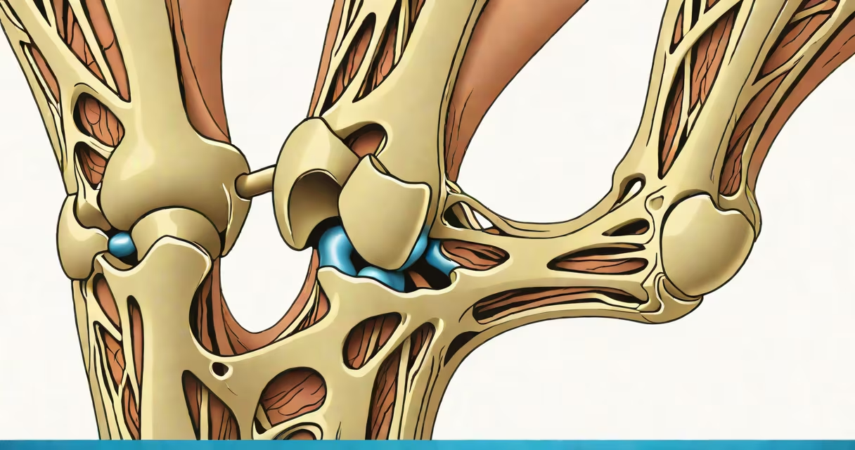
A torn ligament appears as disrupted, frayed, or completely severed connective tissue on imaging scans such as MRI or ultrasound. The damage often shows as increased signal intensity, abnormal tissue texture, or complete absence of continuity in the ligament structure.
Ligaments are dense bands of collagen fibers that stabilize joints by connecting bone to bone. When torn, they lose structural integrity. A partial tear may show as thickened or irregular tissue with fluid accumulation. A complete rupture appears as a gap in the ligament's normal linear pattern, often accompanied by swelling or blood pooling around the joint.
Imaging is essential for accurate diagnosis. On MRI, torn ligaments typically display hyperintense signals on T2-weighted sequences due to edema and hemorrhage. Ultrasound can detect tears in real time, especially in superficial joints like the ankle or wrist. Without imaging, visual inspection cannot confirm a tear-symptoms like pain, swelling, and instability are indicators, not definitive proof.
MRI remains the gold standard for diagnosing ligament injuries. Radiologists assess ligament appearance using signal intensity, morphology, and spatial relationships. A healthy ligament appears as a thin, dark, continuous band on T1 and T2 MRI sequences. A torn ligament shows disruption in this pattern.
For example, an ACL tear in the knee often appears as a discontinuous or thickened ligament with surrounding fluid. A grade 1 tear shows mild swelling and microscopic fiber separation. Grade 2 involves partial tearing with visible gaps. Grade 3 is a complete rupture-no continuous tissue is seen, and the joint becomes unstable.
Ultrasound offers real-time changing assessment. A trained technician can observe ligament movement during joint motion. A torn ligament may fail to glide smoothly or show abnormal displacement under stress. This method is less sensitive than MRI but useful in acute settings where immediate results are needed.
The most frequently injured ligaments occur in weight-bearing and high-motion joints: the knee, ankle, shoulder, and wrist. Each has distinct imaging features.
| Joint | Common Ligament | Appearance on MRI/US |
|---|---|---|
| Knee | Anterior Cruciate Ligament (ACL) | Discontinuity, thickening, or abnormal signal; often associated with bone bruising on adjacent femur or tibia |
| Ankle | Anterior Talofibular Ligament (ATFL) | Thickened, irregular signal; gap or loss of continuity; fluid in joint space |
| Shoulder | Coracoclavicular Ligament | Partial or complete tear visible as a gap between clavicle and coracoid process; often seen in acromioclavicular sprains |
| Wrist | Scapholunate Ligament | Increased signal; widening of the scapholunate interval (>3 mm); "sign of the scaphoid" on axial view |
For instance, a patient with a sudden ankle twist may have an ATFL tear confirmed via ultrasound. The image shows a gap in the ligament's normal linear structure, with fluid accumulation around the lateral malleolus. The joint feels unstable during weight-bearing tests.
Non-imaging clues include joint swelling, bruising, and inability to bear weight. However, these signs overlap with fractures and tendinitis. Only imaging distinguishes a torn ligament from similar conditions.
Many assume a torn ligament causes immediate, severe pain. But pain levels vary widely. Some patients report minimal discomfort despite a complete ACL tear. Others feel intense pain with a minor sprain. Pain is not a reliable indicator of ligament integrity.
Another misconception is that ligaments heal quickly. In reality, ligaments have poor blood supply, limiting their ability to regenerate. Most tears require surgical repair or prolonged rehabilitation. A 2021 study in the Journal of Orthopaedic Research found that 60% of ACL tears in adults do not heal spontaneously without intervention.
Some people mistake ligament injuries for muscle strains. Both cause pain and swelling. But ligaments stabilize joints. A torn ligament leads to joint instability-feeling like the knee gives way or the ankle rolls repeatedly. Muscle strain affects movement but not joint alignment.
I once observed a college athlete who dismissed his ankle injury as a "twist." He returned to play after two days. An MRI revealed a complete ATFL tear with joint effusion. He required six weeks of bracing and physical therapy. Early imaging prevented long-term instability.
If a joint feels unstable, swells rapidly, or fails to support weight within 24 hours of injury, imaging is necessary. Delaying care increases risk of chronic instability and secondary joint damage.
Red flags include:
Even if symptoms improve, a torn ligament may not heal properly. Untreated tears lead to osteoarthritis over time. The American Academy of Orthopaedic Surgeons reports that untreated ACL tears increase the risk of meniscus tears by 50% within five years.
Early diagnosis improves outcomes. If you suspect a ligament injury, consult a healthcare provider. Imaging can confirm the extent of damage and guide treatment-whether conservative management or surgical reconstruction.