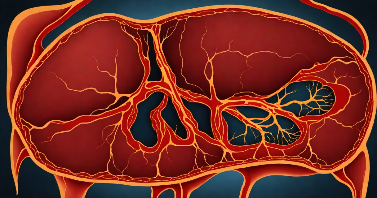
A healthy liver is a dark red-brown organ with a smooth, slightly rubbery texture, measuring roughly 6 inches across and weighing about 3 pounds. It sits under the diaphragm on the right side of the abdomen and plays a central role in filtering toxins, metabolizing nutrients, and producing bile.
The liver's color comes from its high concentration of blood vessels and iron-rich hepatocytes-the primary functional cells. A normal liver appears uniformly pigmented without mottling, nodules, or discoloration. Its surface is smooth, without scars or irregularities. When viewed in cross-section, the tissue shows consistent density and no visible cysts, fibrosis, or fatty deposits.
Medical imaging such as ultrasound or MRI confirms internal structure. A healthy liver appears homogeneous-without areas of increased or decreased echogenicity-and maintains proper vascular flow. Abnormal findings include fatty infiltration (bright spots on ultrasound), cirrhotic scarring (nodular texture), or tumors (distinct masses).
The liver is divided into eight anatomical segments based on blood supply and drainage. Each segment contains lobules-tiny functional units shaped like hexagons. At the center of each lobule is a central vein; radiating outward are portal triads containing a branch of the hepatic artery, portal vein, and bile duct.
Hepatocytes make up 80% of the liver's mass. These cells are packed with mitochondria and enzymes needed for detoxification. Under a microscope, healthy hepatocytes appear round or polygonal with clear cytoplasm and prominent nuclei. There is no fat accumulation, inflammation, or cell death.
When liver function declines, changes occur at the cellular level. Fatty liver disease causes lipid droplets to fill hepatocytes, making them appear foamy under a microscope. Alcoholic hepatitis leads to ballooning degeneration-cells swell and lose their structure. Chronic injury triggers fibrosis, where collagen fibers replace normal tissue, eventually forming scar tissue.
Compare a healthy liver to one affected by disease using real clinical data:
| Feature | Healthy Liver | Diseased Liver (Fatty, Cirrhotic) |
|---|---|---|
| Color | Dark red-brown, uniform | Faint yellow (fatty), grayish (cirrhotic) |
| Texture | Smooth, firm | Nodular, hard (cirrhosis), soft (steatosis) |
| Size | ~6 inches, weighs ~3 lbs | Enlarged (early fatty), shrunken (advanced cirrhosis) |
| Microscopic Appearance | Regular lobules, no fat, no necrosis | Lipid droplets, dead cells, fibrous bands |
According to the American Liver Foundation, fatty liver affects 25% of adults in the U.S., often without symptoms. Early-stage fatty liver may appear normal on imaging but shows microscopic fat accumulation. In contrast, advanced cirrhosis reduces liver volume by up to 30% and increases stiffness-a change detectable via elastography.
Many assume a healthy liver is pale or light-colored because of its association with "clean" organs. This is incorrect. The liver's rich blood supply gives it a deep red-brown hue. A pale liver indicates anemia, severe blood loss, or chronic disease-conditions that impair oxygen delivery to tissues.
Another misconception is that liver size correlates directly with health. A large liver can result from congestion, fatty infiltration, or tumor growth. Conversely, a small liver suggests long-term damage. The National Institutes of Health notes that liver volume below 1,000 mL in adults is associated with poor outcomes in transplant candidates.
During surgery, surgeons assess liver health by touch and visual inspection. A healthy liver feels resilient under pressure and returns to shape quickly. A diseased liver feels brittle or spongy. In one study published in *Hepatology*, surgeons correctly identified early cirrhosis in 87% of cases based on tactile feedback alone.
In a 2022 case reported by Johns Hopkins Medicine, a 54-year-old man with end-stage liver disease underwent evaluation for transplant. His CT scan showed a shrunken, nodular liver with irregular margins and reduced blood flow. Biopsy confirmed bridging fibrosis and regenerative nodules-hallmarks of cirrhosis.
By contrast, a healthy donor liver from a 32-year-old woman had a uniform dark red-brown appearance, smooth surface, and normal vascular architecture. The organ was transplanted successfully, with no rejection episodes over 18 months. Post-op imaging confirmed normal perfusion and absence of scarring.
These cases illustrate that visual and tactile cues remain important-even with advanced imaging. As the Mayo Clinic states, "No scan replaces direct observation during surgery."
A healthy liver is dark red-brown, smooth, firm, and approximately 6 inches wide. It functions optimally when free of fat, scarring, or structural distortion. Any deviation-color change, nodularity, or abnormal texture-signals underlying pathology. Regular screening, especially for those with risk factors like alcohol use or metabolic syndrome, is essential for early detection.