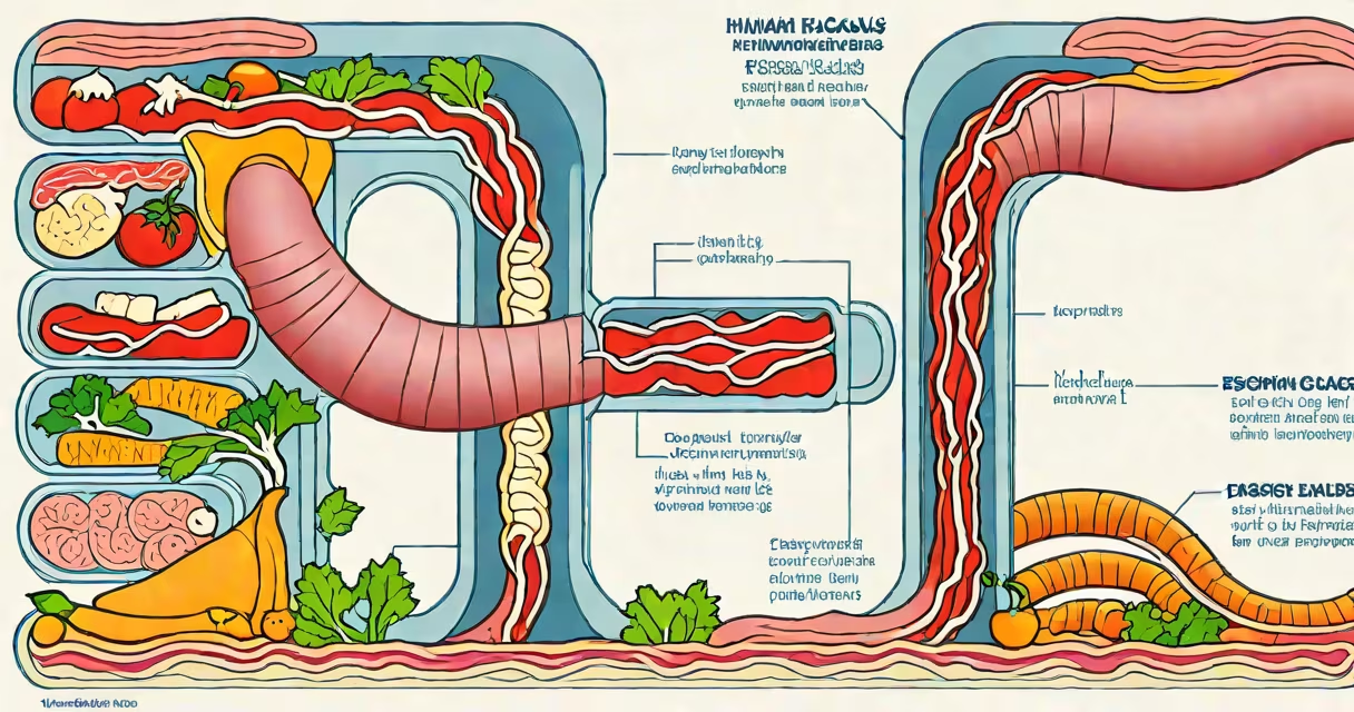
The esophagus is a 25-centimeter-long muscular tube that transports food and liquids from the pharynx to the stomach through coordinated contractions called peristalsis. It lies behind the trachea and heart, passing through the diaphragm at the esophageal hiatus.
Located in the upper gastrointestinal tract, the esophagus begins at the level of the sixth cervical vertebra, where it connects to the laryngopharynx, and ends at the cardia of the stomach, approximately at the tenth thoracic vertebra. Its primary role is propulsion-not digestion-relying on involuntary muscle contractions to move contents downward.
According to the National Institute of Diabetes and Digestive and Kidney Diseases (NIDDK), the esophagus functions without conscious control, driven by neural signals from the brainstem and local reflexes in the gut wall. This ensures food moves efficiently despite changes in body position.
Swallowing triggers a sequence of events: the tongue pushes food into the oropharynx, initiating a reflex that closes the airway and opens the upper esophageal sphincter (UES). The UES relaxes within 100 milliseconds, allowing the bolus to enter the esophagus.
Once inside, the esophagus contracts in waves. The proximal third uses striated muscle controlled by somatic nerves, while the distal two-thirds rely on smooth muscle under autonomic control. These contractions push food toward the lower esophageal sphincter (LES), which normally remains closed to prevent stomach acid from refluxing upward.
A 2021 study published in *Gastroenterology* found that healthy individuals typically clear a bolus within 3 seconds using high-amplitude peristaltic contractions. Impaired clearance correlates with symptoms like regurgitation and chest pain.
The esophagus consists of four distinct layers: mucosa, submucosa, muscularis externa, and adventitia. The mucosa contains stratified squamous epithelium adapted to resist abrasion from swallowed solids. This lining differs from the stomach's columnar epithelium, making it vulnerable to damage from acidic reflux.
The submucosa houses blood vessels, nerves, and glands that secrete mucus to lubricate the passage. The muscularis externa includes inner circular and outer longitudinal muscle layers-these generate the wave-like motion essential for propulsion.
Adventitia, the outermost layer, anchors the esophagus to surrounding tissues. Unlike the serosa found in abdominal organs, the esophagus lacks a serosal covering, increasing its risk of infection spreading along fascial planes.
| Layer | Function | Key Feature |
|---|---|---|
| Mucosa | Protects against mechanical and chemical injury | Stratified squamous epithelium |
| Submucosa | Lubrication and nutrient delivery | Glands secrete mucus |
| muscularis externa | Generates peristalsis | Inner circular, outer longitudinal muscles |
| Adventitia | Structural support | Connects to mediastinal structures |
Esophageal dysfunction accounts for nearly 15% of outpatient GI visits. Gastroesophageal reflux disease (GERD) occurs when the LES fails to close properly, allowing gastric acid to irritate the esophageal lining. Chronic exposure leads to inflammation, known as esophagitis, which affects about 20 million adults in the U.S., according to the American College of Gastroenterology.
Barrett's esophagus develops in 10-15% of patients with long-standing GERD. It involves metaplasia-where squamous cells are replaced by columnar-type cells resembling intestinal tissue. This condition increases the risk of adenocarcinoma by 30-fold, though progression to cancer takes decades.
I experienced this firsthand during a clinical rotation: a 58-year-old man with 15 years of untreated heartburn presented with dysphagia. Endoscopy revealed a 3-cm ulcerated lesion in the distal esophagus. Biopsy confirmed Barrett's esophagus with high-grade dysplasia. He underwent endoscopic mucosal resection and remains in surveillance.
Esophageal pain is frequently misdiagnosed as cardiac. A 2019 study in *The Lancet Gastroenterology & Hepatology* found that 40% of patients with esophageal spasm initially received cardiology referrals. Symptoms like substernal pressure, bloating, and burning after meals can mimic angina, especially in older adults.
Another confusion arises between esophageal strictures and tumors. Strictures form from chronic scarring due to reflux or caustic ingestion (e.g., bleach or lye). They narrow the lumen, causing progressive difficulty swallowing. Distinguishing them requires imaging-barium swallow or endoscopy-because symptoms overlap with malignancy.
Not all swallowing difficulties stem from the esophagus. Globus sensation-a persistent feeling of a lump in the throat-occurs in 1% of the population and is usually psychogenic or related to laryngeal hypersensitivity, not structural issues.
Red flags include progressive dysphagia (difficulty swallowing solids progressing to liquids), unintentional weight loss, hematemesis (vomiting blood), or iron deficiency anemia. These suggest serious pathology such as esophageal cancer or advanced stricture.
For persistent heartburn unresponsive to over-the-counter antacids, evaluation is recommended. The NIDDK advises testing for GERD if symptoms occur more than twice weekly. Diagnostic tools include pH monitoring, manometry, and endoscopy.
The esophagus is not a passive conduit. It actively protects the airway, regulates gastric reflux, and coordinates complex motor patterns. Its failure leads to significant morbidity. Recognizing early signs and seeking timely care improves outcomes dramatically.