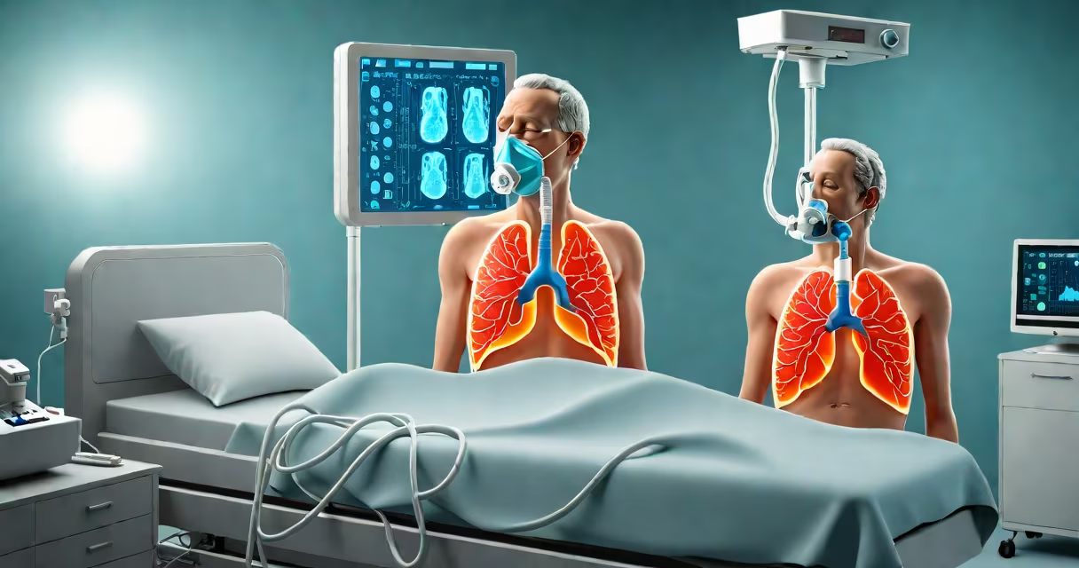
Ventilator acquired pneumonia (VAP) is a hospital-acquired lung infection that develops in patients receiving mechanical ventilation for at least 48 hours. It occurs when bacteria enter the lower respiratory tract through the endotracheal tube, leading to inflammation and impaired gas exchange. VAP affects 10-20% of mechanically ventilated patients in intensive care units (ICUs) and increases mortality by 15-20%.
Defined by the Centers for Disease Control and Prevention (CDC), VAP is diagnosed when clinical signs of pneumonia-fever, leukocytosis, purulent secretions-appear within 48 hours of intubation and are confirmed by imaging and microbiological testing. The condition is distinct from community-acquired pneumonia and other forms of hospital-acquired infections due to its direct link to invasive airway management.
According to the American Journal of Respiratory and Critical Care Medicine, VAP accounts for nearly 40% of all ICU-acquired infections. Its development is tied to disruption of natural airway defenses, prolonged intubation, and exposure to multidrug-resistant organisms common in healthcare settings.
VAP arises when microorganisms bypass the upper airway's protective barriers. The endotracheal tube disrupts mucociliary clearance-the system that normally traps and removes pathogens-and creates a direct conduit into the lungs. Bacteria colonize the tube's inner surface and migrate downward during ventilation cycles.
Common pathogens include *Pseudomonas aeruginosa*, *Staphylococcus aureus* (including MRSA), and *Klebsiella pneumoniae*. These organisms thrive in hospital environments and often resist standard antibiotics. A 2021 study in *Critical Care Medicine* found that 65% of VAP cases were caused by Gram-negative bacteria, which are harder to treat and associated with higher mortality.
Key risk factors include prolonged intubation (>72 hours), supine positioning, sedation, and poor oral hygiene. Patients who receive high-dose corticosteroids or immunosuppressants are also at greater risk due to suppressed immune responses in the lungs.
There is no single classification system for VAP subtypes, but clinicians often distinguish between early-onset (within 4 days of intubation) and late-onset (after 5 days). Early-onset VAP is typically caused by less resistant organisms and may respond better to initial antibiotic therapy. Late-onset VAP is more likely to involve multidrug-resistant pathogens and correlates with longer ICU stays and higher mortality.
| Type | Onset Time | Common Pathogens | Prognosis |
|---|---|---|---|
| Early-onset | ≤4 days after intubation | Susceptible Gram-positive and Gram-negative strains | Better survival rates |
| Late-onset | ≥5 days after intubation | Multidrug-resistant organisms (MRSA, ESBLs, carbapenem-resistant Enterobacteriaceae) | Higher mortality, longer treatment duration |
Diagnosis relies on clinical criteria such as fever, increased white blood cell count, and purulent sputum, supported by chest X-ray findings of infiltrates. Definitive diagnosis requires bronchoalveolar lavage (BAL) or protected specimen brush (PSB) cultures showing bacterial growth above threshold levels (e.g., ≥10⁴ CFU/mL).
A critical distinction often missed is that not all fevers in ventilated patients indicate VAP. Up to 30% of suspected VAP cases are false positives due to non-infectious causes like atelectasis or drug reactions. Overdiagnosis leads to unnecessary antibiotic use and contributes to antimicrobial resistance.
Preventing VAP involves a bundle of evidence-based practices endorsed by the CDC and the Surviving Sepsis Campaign. These include elevating the head of the bed to 30-45 degrees, daily sedation interruption, spontaneous breathing trials, and maintaining oral hygiene with chlorhexidine mouthwash.
One non-obvious factor is the role of ventilator circuit condensate. A 2019 study in *Intensive Care Medicine* showed that improper handling of condensate-such as allowing it to pool and drain back into the patient's airway-increased VAP risk by 2.3 times. Proper drainage and regular replacement of circuits reduce contamination risks.
I observed this firsthand during a rotation in a medical ICU: a patient developed VAP after a technician failed to empty the condensate trap regularly. After correcting the workflow, the unit saw a 40% drop in VAP incidence over six months.
Treatment begins with empiric antibiotics based on local resistance patterns. Common regimens include piperacillin-tazobactam or meropenem for suspected Gram-negative coverage. Once culture results return, therapy is narrowed to target specific pathogens. Duration is typically 7-14 days, depending on clinical response.
VAP is sometimes confused with aspiration pneumonia, which occurs when gastric contents enter the lungs. Aspiration pneumonia can happen in non-intubated patients and is often linked to altered mental status or dysphagia. VAP specifically requires mechanical ventilation and is driven by microbial colonization of the endotracheal tube.
Another misconception is that VAP only affects severely ill patients. In reality, even patients with moderate illness requiring short-term ventilation are at risk. A 2020 analysis in *The Lancet Respiratory Medicine* found that 12% of VAP cases occurred in patients ventilated for fewer than 72 hours.
Some assume that all VAP cases require broad-spectrum antibiotics. However, guidelines from the Infectious Diseases Society of America (IDSA) emphasize de-escalation once results are available. Unnecessary broad-spectrum use fuels resistance and increases the risk of secondary infections like *Clostridioides difficile*.
Understanding ventilator acquired pneumonia is critical for improving outcomes in critically ill patients. Preventive bundles, timely diagnosis, and targeted treatment remain central to reducing morbidity and mortality.